A 71-year-old asymptomatic woman with severe coronary artery disease and a history of coronary artery bypass grafting (CABG) 15 years ago underwent contrast-enhanced computed tomography of the chest for tumour staging in colorectal carcinoma. As an incidental finding, she was noted to have a mostly thrombosed, huge aneurysm of the saphenous vein graft (SVG) to the left anterior descending artery (LAD) (Figure 1A and 1B), maximum diameter 106 mm. Coronary angiography demonstrated a perfused lumen of the partially thrombosed SVG aneurysm with a filiform stenosis directly distal to the vein graft aneurysm (Figure 1C, Moving image 1). Furthermore, aneurysms of the left main coronary artery (Figure 1D, Moving image 2), the proximal LAD and the chronically occluded proximal right coronary artery were found. Unfortunately, the patient denied any cardiac surgery or percutaneous coronary intervention of her aneurysm and passed away shortly after discharge from hospital. An autopsy was declined by the patient’s family.
The SVG to the LAD is the most common site for an aneurysm formation, followed by the right coronary artery, and least commonly, the left circumflex. SVG aneurysms usually become evident 10-15 years after CABG. The incidence of significant SVG aneurysm (0.1%) is probably underestimated because the initial presentation may be rupture leading to sudden death, the aneurysm may not appear on angiography if it contains significant thrombus, and many patients – as ours – are asymptomatic.1,2
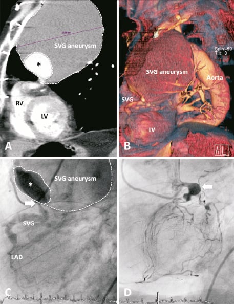
Figure 1. A) CT angiogram of the mostly thrombosed saphenous vein graft (SVG) aneurysm (maximum diameter 106 mm) with a small perfused lumen (*). RV: right ventricle; LV: left ventricle. B) Three-dimensional CT reconstruction of the SVG aneurysm in LAO 60° view. C) Angiogram of the SVG with a filiform stenosis (white arrow) distal to the huge SVG aneurysm in LAO 60° view. D) Coronary angiogram of the left coronary artery with aneurysms of the left main coronary artery (maximum diameter 21 mm, white arrow) and the ostial left anterior descending artery in LAO 60° view.
Online data supplement
Video 1. LAO 60° view of the saphenous vein graft (SVG) with a filiform stenosis distal to the huge SVG aneurysm.
Video 2. LAO 60° view of the left coronary artery with aneurysms of the left main coronary artery (maximum diameter 21 mm) and the ostial left anterior descending artery.