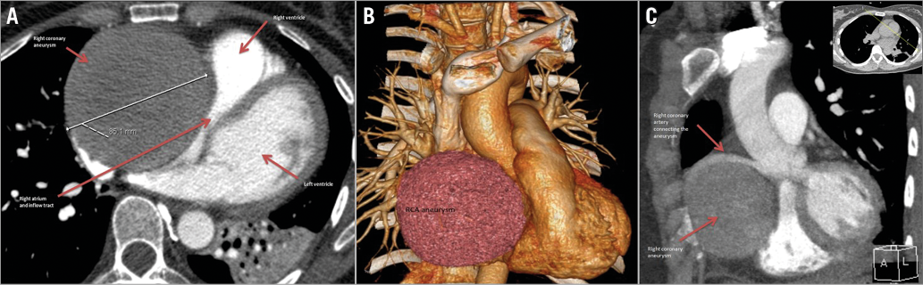
A 32-year-old healthy woman presented with impaired exercise tolerance four months after giving birth to her fourth child. Chest radiography showed increased contour size of the right atrium. Computed tomographic angiography (CTA) was arranged with the initial intention of excluding pulmonary embolism. A large ball-shaped structure, 8 cm in diameter, was found across the heart in the right-hand side of the arterioventricular groove with significant compression of the right atrium, the right ventricle and the superior vena cava. It was initially suspected to be a huge pericardial cyst (Panel A, Panel B). Echocardiography showed a large round structure (8×8.5 cm) consisting of turbulent flow (Moving image 1). A coronary angiogram confirmed the structure was a huge right coronary aneurysm with turbulent flow (Moving image 2, Moving image 3). Urgent coronary artery bypass grafting with a saphenous venous graft was performed. The aneurysm was resected. Preoperatively, CTA was repeated: it showed that there was connection of the proximal right coronary artery with the aneurysm, signifying that this was a coronary aneurysm and not a pericardial cyst (Panel C).
The patient’s symptoms improved after the operation. Common causes for coronary aneurysm are or include vasculitis, connective tissue disorders, mycotic, congenital, atherosclerosis and idiopathic. Histological assessment of the resected right coronary artery showed slight mucoid degeneration of the media, without evidence of pronounced atherosclerosis or signs of vasculitis. The largest dimensions of the aneurysm were 8.5×3.5×0.2 cm.
This case is an example of a huge idiopathic coronary aneurysm. The presentation was vague. It is uncommon for a coronary aneurysm to develop to such a large size without a cardiac event.
Conflict of interest statement
The authors have no conflicts of interest to declare.
Supplementary data
Moving image 1. Echocardiography showing large round structure with turbulent flow.
Moving image 2. Coronary angiogram showing huge coronary aneurysm.
Moving image 3. Coronary angiogram confirming huge coronary aneurysm.
Supplementary data
To read the full content of this article, please download the PDF.
Echocardiography showing large round structure with turbulent flow.
Coronary angiogram showing huge coronary aneurysm.
Coronary angiogram confirming huge coronary aneurysm.