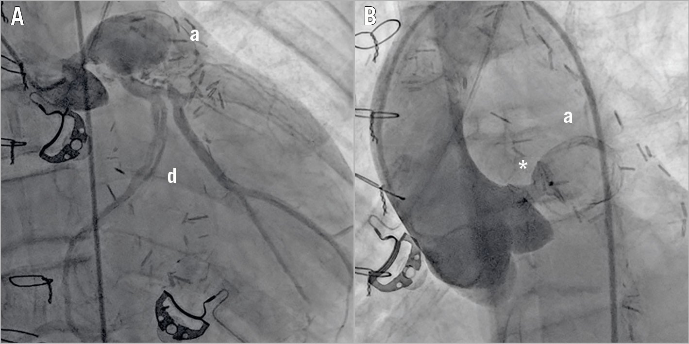

Figure 1. Fluoroscopic images. A) Coronary angiogram showing the aneurysm (a) and dissection (d). B) Aortogram showing the device (*) deployed.
Chronic coronary aneurysms are present in 6% of patients with Kawasaki disease (KD) and are associated with long-term morbidity and mortality1,2. There are several interventional treatment options for coronary aneurysms3. Herein we report the first successful use of transcatheter occlusion of a huge left main coronary artery (LMCA) aneurysm due to KD with an AMPLATZER™ Muscular Ventricular Septal Occluder device (VSO; St. Jude Medical, St. Paul, MN, USA).
The 16-year-old male patient with previous history of KD and coronary artery bypass grafting presented with exercise-induced ventricular tachycardia (VT). Baseline echocardiogram (ECG) and laboratory tests were unremarkable. The coronary angiogram showed a 4 cm diameter left main coronary artery (LMCA) aneurysm, patent grafts to the left anterior descending (LAD) and left circumflex (LCX) arteries and dissection in the posterolateral (PL) branch of the LCX (Figure 1A, Moving image 1). Following implantable cardioverter defibrillator (ICD) implantation, transcatheter closure of the LMCA was planned. After right femoral artery access and administration of 100 IU/kg of unfractionated heparin, two hydrophilic coronary guidewires were positioned in the LCX and the delivery catheter was positioned in the LMCA. A 6 mm AMPLATZER Muscular VSO was advanced into the aneurysm. The first disc was deployed in the isthmus of the aneurysm and the second disc was deployed in the aortic side of the LMCA ostium. The patient was observed for 15 minutes before release of the device (Figure 1B, Moving image 2, Moving image 3). The ostia of the LAD and LCX were not affected, as shown by non-selective visualisation during aortography. Clopidogrel was discontinued following dual antiplatelet therapy for 12 months.
We demonstrated that transcatheter occlusion of ostial coronary aneurysms with occlusion devices is a safe and efficient method for protecting patients from complications in the long run.
Conflict of interest statement
The authors have no conflicts of interest to declare.
Supplementary data
To read the full content of this article, please download the PDF.
Moving image 1. Coronary angiogram showing the aneurysm and the dissection.
Moving image 2. Aortogram before the device was released.
Moving image 3. Aortogram after the release of the device, small through-device leak.

