DOI: 10.4244/EIJV8I6A116

Giant coronary aneurysm in Churg-Strauss syndrome

Marc Hartmann1*, MD, PhD; Elly M.C.J. Wajon1, MD; Gert K. van Houwelingen1, MD; Martin G. Stoel1, MD; Clemens von Birgelen1,2, MD, PhD

A 64-year-old male with Churg-Strauss syndrome (CSS) presented with worsening dyspnoea without chest pain besides having long-standing asthma. There were no cardiovascular risk factors. The electrocardiogram showed signs of prior anterior infarction. Echocardiography demonstrated severely depressed left ventricular function (ejection fraction 30%). Multislice cardiac computed tomography revealed a giant coronary aneurysm (Figure 1A and Figure 1B) of the left circumflex coronary (LCX) artery of 4 cm diameter with mural thrombi (Figure 1B) and also aneurysmatic changes of the right coronary artery (RCA) with proximal occlusion (Figure 1 A). Coronary angiography showed aneurysmatic changes of the LCX with collateral filling of distal RCA and mid-segment occlusion of the left anterior descending (LAD) (online Figure 1C and 1D, Moving image 1 - Moving image 3). Single-photon emission tomography confirmed an ischaemic cardiomyopathy with irreversible defects. Treatment was carried out medically, and an internal cardioverter-defibrillator was implanted.
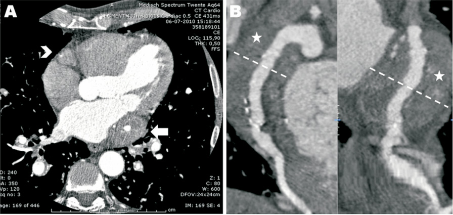
Figure 1. Multislice cardiac computed tomography imaging (A, B) showed a giant coronary aneurysm of the LCX artery (arrow in A) with a diameter of 4 cm and mural thrombi (asterisk in B) and aneurysmatic changes of the RCA with proximal occlusion (arrowhead in A).

Coronary aneurysms are uncommon; they are usually due to atherosclerotic disease and only in a minority of cases are they the result of an autoimmune vasculitis1. CSS is a rare small-vessel vasculitis with asthma and eosinophilia2,3. There are only a few reports with CSS and involvement of major coronary vessels with aneurysmatic changes1-3. The toxic effects of eosinophils on the coronary wall may result in aneurysmatic enlargement1-3. This case describes extensive coronary involvement leading to an ischaemic cardiomyopathy, probably due to thrombotic occlusions and distal coronary embolisations1.

Conflict of interest statement

The authors have no conflict of interest to declare.

Online data supplement

Figure 1 C and Figure 1 D. Coronary angiography (with CLS guiding catheter) also showed extensive aneurysmatic changes of the LCX artery (arrow in C, D) with collateral filling of distal RCA which is proximally occluded (arrowhead in C, D). The LAD showed mid-segment occlusion with minor collateral filling of the distal part (asterisk in C, D).

Moving image 1. Coronary angiogram RCA.

Moving image 2. Coronary angiogram LCA#1.

Moving image 3. Coronary angiogram LCA#2.
Volume 8 Number 6
Oct 26, 2012
Volume 8 Number 6
View full issue


Key metrics

Suggested by Cory

IMAGE IN CARDIOLOGY

10.4244/EIJV12I9A190 Oct 20, 2016
An unexpected huge coronary aneurysm
Chan Y et al
free

IMAGE IN CARDIOLOGY

10.4244/EIJV12I1A11 May 16, 2016
Embolic myocardial infarction due to coronary artery aneurysm in a patient with Loeys-Dietz syndrome
Cereda A et al
free

IMAGE IN CARDIOLOGY

10.4244/EIJ-D-16-00393 Apr 20, 2017
Multiple spontaneous intimal dissections with single left coronary artery and coronary artery aneurysm
Achkouty G et al
free

IMAGE IN CARDIOLOGY

10.4244/EIJ-D-16-00316 Jun 2, 2017
Inflammatory coronary ectasia identified by three-dimensional volume rendering of 18F-Fluorodeoxyglucose PET/CT
Park E et al
free

10.4244/EIJV11I7A171 Nov 20, 2015
How should I treat multiple coronary aneurysms with severe stenoses?
Warisawa T et al
free

10.4244/EIJV9I11A228 Mar 20, 2014
How should I treat a coronary bifurcation wide-neck aneurysm?
Sgueglia G et al
free
Trending articles
334.7

State-of-the-Art Review

10.4244/EIJ-D-21-00904 Apr 1, 2022
Antiplatelet therapy after percutaneous coronary intervention
Angiolillo D et al
free
84.05

State-of-the-Art

10.4244/EIJ-D-23-00840 Sep 2, 2024
Aortic regurgitation: from mechanisms to management
Baumbach A et al
free
45.75

State-of-the-Art

10.4244/EIJ-D-25-01346 May 4, 2026
Slow flow and no reflow after percutaneous coronary intervention
Brugaletta S et al
free
42.15

State-of-the-Art

10.4244/EIJ-D-25-00896 Apr 6, 2026
Pretreatment with antiplatelet agents in patients undergoing coronary revascularisation
Kaur G et al
free
38.2

State-of-the-Art

10.4244/EIJ-D-24-00195 Apr 7, 2025
Percutaneous coronary intervention for calcified and resistant lesions
Pesarini G et al
free
33.5

Original Research

10.4244/EIJ-D-26-00032 May 15, 2026
Glucocorticoids to reduce permanent pacemaker implantation after TAVI: the GLUCO-TAVI randomised trial
Fuertes-Kenneally L et al
32.2

State-of-the-Art

10.4244/EIJ-D-25-00874 Jun 1, 2026
TAVI and coronary interventions: indications, technical considerations, and clinical scenarios
Aquino Bruno H et al
free
29.2

Original Research

10.4244/EIJ-D-25-01370 May 21, 2026
Prognostic value of early haemodynamic valve deterioration after TAVI
Trimaille A et al
29.2

Original Research

10.4244/EIJ-D-25-01370 Jun 1, 2026
Prognostic value of early haemodynamic valve deterioration after TAVI
Trimaille A et al
X

PCR
Impact factor: 9.2
2025 Journal Citation Reports®
Science Edition (Clarivate Analytics, 2026)
Online ISSN 1969-6213 - Print ISSN 1774-024X
© 2005-2026 Europa Group - All rights reserved