![]()
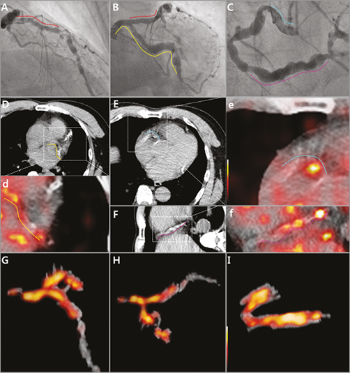
A 59-year-old man was admitted to our hospital complaining of worsening exertional chest pain. His coronary angiography revealed diffuse coronary artery ectasia (CAE) accompanied by multiple severe stenotic lesions across all of the three vessels (Panel A-Panel C, Moving image 1). After preparing to suppress 18F-Fluorodeoxyglucose (FDG) uptake of myocardium (Appendix Figure 1), we performed FDG PET imaging with CT co-registration to estimate the inflammatory process of his coronary ectasia. While the fused PET/CT images demonstrated FDG-avid coronary artery ectasia, we processed PET signals through thresholding background noise using MATLAB R2014b software (The MathWorks, Natick, MA, USA) (Panel D-d, Panel E-e, Panel F-f) to acquire optimal localisation of the vascular FDG uptake in the coronary trees. Then, we produced a three-dimensional rendering of the coronary arterial images from the co-registered PET/CT images using ImageJ (NIH, Bethesda, MD, USA) and OsiriX software (The OsiriX Foundation, Geneva, Switzerland) (Panel G, Panel H, Panel I, Moving image 2). After superimposing the PET signals on the coronary masks generated by segmentation on the basis of anatomical CT morphology, we reconstructed three-dimensional (3D) coronary PET/CT (Appendix Figure 2). The patient underwent bypass surgery with prescription of dual antiplatelet agents, and high-dose statin.
In conclusion, current 3D reconstruction PET/CT of coronary artery trees demonstrated the increased metabolic activity of FDG precisely matched with coronary ectatic segments. While it is still unclear whether the patient’s ectatic lesion originated from systemic vasculitis or solely an atherosclerotic process, the underlying pathogenesis of CAE could be related to active inflammation of the coronary arterial walls.
Funding
This research was supported in part by grants from the National Research Foundation of Korea (NRF), the Ministry of Education, Science and Technology (grant number 2015R1A2A2A07027863).
Conflict of interest statement
The authors have no conflicts of interest to declare.
Supplementary data
Moving image 1. The patient’s initial coronary angiogram. A, B and C are images of the left coronary artery. A) Left anterior oblique view. B) Anterior-posterior caudal view. C) Anterior-posterior cranial view. D) Right coronary artery.
Moving image 2. The rotational 3D reconstructed PET/CT fusion of coronary artery trees. A) Left anterior descending artery. B) Left circumflex artery. C) Right coronary artery.
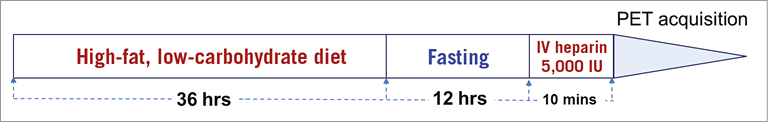
Appendix Figure 1. Patient preparation. The patient initiated a high-fat low-carbohydrate meal 48 hours prior to PET imaging followed by 12-hour fasting, and was administered IV heparin 5,000 IU 10 minutes before the examination.
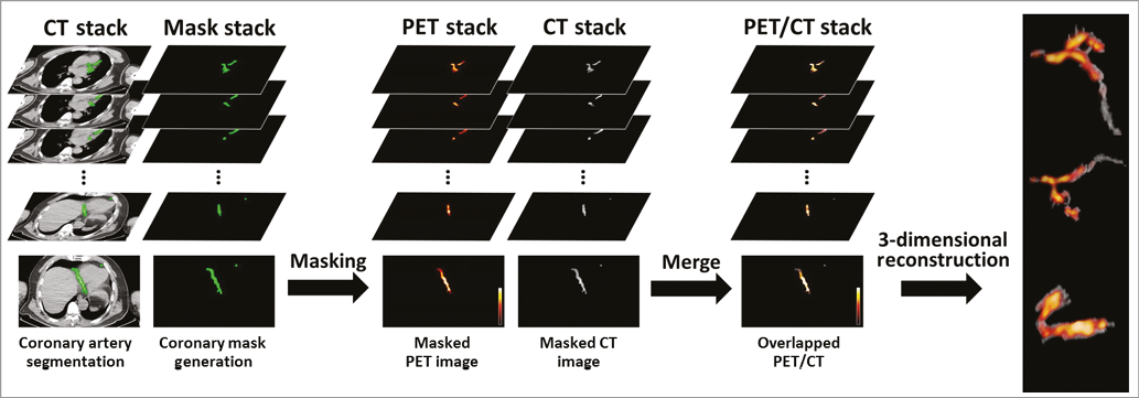
Appendix Figure 2. Coronary masking and subsequent 3D imaging reconstruction process. The segments of coronary arteries in the CT stack were identified using an image processing program, ImageJ (NIH, Bethesda, MD, USA). The segmentation process was performed just on the basis of anatomical morphology of CT without the PET information. PET images were masked with the coronary masks, which were generated by the segmentation process. After superimposing the PET signals on the CT images produced by CT coronary segmentation, we reconstructed 3-dimensional images of PET/CT using a DICOM viewer, OsiriX (The OsiriX Foundation, Geneva, Switzerland).
Supplementary data
To read the full content of this article, please download the PDF.
1A. The patient’s initial coronary angiogram.
1B. The patient’s initial coronary angiogram.
1C. The patient’s initial coronary angiogram.
1D. The patient’s initial coronary angiogram.
2A. The rotational 3D reconstructed PET/CT fusion of coronary artery trees.
2B. The rotational 3D reconstructed PET/CT fusion of coronary artery trees.
2C. The rotational 3D reconstructed PET/CT fusion of coronary artery trees.