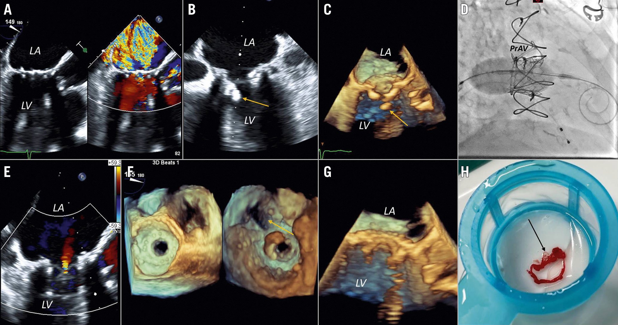
Figure 1. Intraprocedural imaging for transcatheter mitral valve-in-valve implantation. A) Transoesophageal echocardiogram (TOE) (mid-oesophageal long axis view with colour Doppler comparison) demonstrating severe mitral regurgitation (MR). B) TOE (mid-oesophageal four-chamber view) demonstrating flail mitral leaflet material (arrow). C) TOE (mid-oesophageal long axis three-dimensional view) demonstrating flail mitral leaflet material (arrow). D) Fluoroscopy-guided mitral valve in valve deployment. E) TOE (mid-oesophageal long axis view with colour Doppler) demonstrating mild transvalvular MR. F) Dual layout 3D view of the mitral valve demonstrating mitral valve in valve without left ventricular outflow tract obstruction (arrow). G) TOE (mid-oesophageal long axis 3D view) of the mitral valve-in-valve. H) Leaflet fragment of the bioprosthetic valve (arrow) captured by the SENTINEL Cerebral Protection System. LA: left atrium; LV: left ventricle; PrAV: prosthetic aortic valve
A 76-year-old woman with a history of concomitant, surgical, bioprosthetic aortic valve (21 mm PERIMOUNT Magna Ease; Edwards Lifesciences) and mitral valve (27 mm Hancock II; Medtronic) replacement five years earlier, due to rheumatic heart disease, presented with acute heart failure decompensation. Three years after surgery she had infective endocarditis, with small vegetations (3-4 mm) attached to the mitral valve, that was treated successfully with antibiotic therapy without residual valvular dysfunction. Two years later, she developed severe symptomatic mitral regurgitation (MR) without evidence of active infection. Due to her comorbidities, including the need for reoperation, severe obstructive lung disease and high Society of Thoracic Surgeons risk score (7.48%), the Heart Team deemed her at high risk and recommended valve-in-valve transcatheter mitral valve implantation. Intraprocedural transoesophageal echocardiogram (TOE) showed severe MR (Figure 1A, Moving image 1) due to a flail leaflet of the bioprosthetic valve (Figure 1B, Figure 1C, Moving image 2, Moving image 3). After transseptal puncture and balloon dilatation were performed under fluoroscopic and TOE guidance, a 26 mm SAPIEN 3 transcatheter heart valve (Edwards Lifesciences) was advanced into the mitral valve, and then positioned and deployed under rapid ventricular pacing (Figure 1D, Moving image 4). After valve deployment, TOE showed mild transvalvular MR (Figure 1E, Moving image 5) without paravalvular leak. The final mean transmitral gradient was 7 mmHg without left ventricular outflow obstruction (Figure 1F). However, the leaflet fragment that was initially noted (Figure 1B, Figure 1C) was no longer seen after the valve deployment (Figure 1E-Figure 1G, Moving image 6). The SENTINEL Cerebral Protection System (Boston Scientific) was used during the procedure and captured large embolic leaflet material that prevented cerebral embolism (Figure 1H). The post-procedural course was uneventful with no neurological deficits.
Conflict of interest statement
The authors have no conflicts of interest to declare.
Supplementary data
To read the full content of this article, please download the PDF.
Moving image 1. TOE (mid-oesophageal long axis view with colour Doppler comparison) demonstrating severe MR.
Moving image 2. TOE (mid-oesophageal four-chamber view) demonstrating flail mitral leaflet material.
Moving image 3. TOE (mid-oesophageal long axis 3D view) demonstrating flail mitral leaflet material.
Moving image 4. Fluoroscopy-guided valve in valve deployment.
Moving image 5. TOE (mid-oesophageal long axis view with colour Doppler comparison) demonstrating mild transvalvular MR.
Moving image 6. TOE (mid-oesophageal long axis 3D view) demonstrating the mitral valve in valve.

