IMAGE IN CARDIOLOGY

DOI: 10.4244/EIJV11I8A194

Complete section of anterior mitral valve during percutaneous valvuloplasty

Amparo Benedicto1*, MD; Guillermo Reyes2, MD; Rio Aguilar1, MD; Fernando Alfonso1, MD

A 35-year-old female was referred for mitral percutaneous valvuloplasty due to a symptomatic rheumatic lesion (severe stenosis with 15 mmHg mean gradient, mild to moderate regurgitation and a Wilkins score of 6).

The basal haemodynamic layout recorded a 23 mmHg mean gradient and a 55 mmHg “v” wave, even if on angiography the regurgitation was quantified as moderate. What drew our attention was an image in the diaphragmatic left ventricular side, originating in the valvular plane, suggesting a severe shortening and chordal fusion (Figure 1A).

A 26 mm Inoue-Balloon Catheter (Toray, Tokyo, Japan) was employed. The post-valvuloplasty angiogram showed massive mitral regurgitation.

Transthoracic (TTE) (Figure 1B, Moving image 1) and transoesophageal (Figure 1C, Moving image 2, Moving image 3) echocardiography (TEE) revealed the complete section of the anterior mitral leaflet, from the free edge to the ring with massive mitral regurgitation (Appendix Figure 1). The valve was severely thickened with both commissures fused.

Urgent surgery was performed. The direct broken valve inspection supported the TEE findings, consistent with severe rheumatic damage both to the leaflet and to the subvalvular apparatus (Figure 1D).

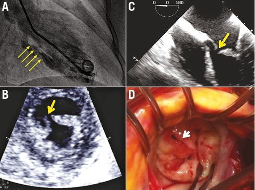
Figure 1. Mitral valve angiographic, echocardiographic and direct views. A) Ventriculography suggesting a severe shortening and chordal fusion. B) Short-axis TTE: section from free edge to the ring. C) Four-chamber TEE: section in the thickened anterior mitral leaflet. D) Surgical view, correlating with echocardiographic findings.

Mitral percutaneous valvuloplasty is supported by years of experience, a previous proper assessment of the involvement of each valvular element being crucial.

Conflict of interest statement

The authors have no conflicts of interest to declare.

Supplementary data

Moving image 1. Complete anterior leaflet section in the short-axis TTE view.

Moving image 2. Complete anterior leaflet section in the long-axis TEE view.

Moving image 3. Massive acute mitral regurgitation in the TEE.

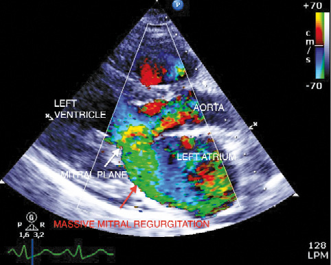
Appendix Figure 1. Massive mitral regurgitation in the long parasternal view.

Supplementary data

To read the full content of this article, please download the PDF.

Moving image 1. Complete anterior leaflet section in the short-axis TTE view.

Moving image 2. Complete anterior leaflet section in the long-axis TEE view.

Moving image 3. Massive acute mitral regurgitation in the TEE.

Volume 11 Number 8
Dec 20, 2015
Volume 11 Number 8
View full issue


Key metrics

Suggested by Cory

Image – Interventional flashlight

10.4244/EIJ-D-20-00325 Sep 20, 2021
A novel technique for percutaneous mitral balloon valvuloplasty
Aurigemma C et al
free

10.4244/EIJV11SWA8 Sep 17, 2015
Mitral valve anatomy: pre-procedural screening and imaging techniques
González-Gómez A et al
free

IMAGE IN CARDIOLOGY

10.4244/EIJY14M07_05 Jul 20, 2015
MitraClip repair of a “trileaflet” regurgitant mitral valve
Freixa X et al
free

Image – Interventional flashlight

10.4244/EIJ-D-18-00973 Apr 16, 2019
Percutaneous repair of healed endocarditis of the mitral valve using MitraClip devices around a large mobile vegetation
Ninios V et al
free

Image – Interventional flashlight

10.4244/EIJ-D-18-00556 Dec 20, 2018
First experience with the MitraClip XTR device for extensive mitral valve prolapse (Barlow’s disease)
Weinmann K et al
free
Trending articles
200.45

State-of-the-Art

10.4244/EIJ-D-21-00089 Jun 11, 2021
Intracoronary optical coherence tomography: state of the art and future directions
Ali ZA et al
free
154.43

State-of-the-Art

10.4244/EIJ-D-22-00776 Apr 3, 2023
Computed tomographic angiography in coronary artery disease
Serruys PW et al
free
92.95

State-of-the-Art Review

10.4244/EIJ-D-20-01296 Aug 27, 2021
Management of cardiogenic shock
Thiele H et al
free
47.4

State-of-the-Art

10.4244/EIJ-D-24-00386 Feb 3, 2025
Mechanical circulatory support for complex, high-risk percutaneous coronary intervention
Ferro E et al
free
43.65

Clinical research

10.4244/EIJ-D-23-00590 Dec 4, 2023
Prognostic impact of cardiac damage staging classification in each aortic stenosis subtype undergoing TAVI
Nakase M et al
free
36.5

State-of-the-Art

10.4244/EIJ-D-23-00448 Jan 15, 2024
Coronary spasm and vasomotor dysfunction as a cause of MINOCA
Yaker ZS et al
free
34.75

State-of-the-Art

10.4244/EIJ-D-23-00606 Jan 1, 2024
Targeting inflammation in atherosclerosis: overview, strategy and directions
Waksman R et al
free
X

The Official Journal of EuroPCR and the European Association of Percutaneous Cardiovascular Interventions (EAPCI)

EuroPCR EAPCI
PCR ESC
Impact factor: 7.6
2023 Journal Citation Reports®
Science Edition (Clarivate Analytics, 2024)
Online ISSN 1969-6213 - Print ISSN 1774-024X
© 2005-2025 Europa Group - All rights reserved