IMAGE IN CARDIOLOGY

DOI: 10.4244/EIJV11I3A62

Calcified plaque ablated by rotational atherectomy visualised by optical coherence tomography

Terumasa Koyama*, MD; Hiroyuki Okura, MD; Teruyoshi Kume, MD; Kenzo Fukuhara, MD; Yoji Neishi, MD; Akihiro Hayashida, MD; Takahiro Kawamoto, MD; Kiyoshi Yoshida, MD

Calcification is visualised by optical coherence tomography (OCT) as a well-delineated, signal-poor region with sharp borders.

A 63-year-old female patient with stable angina underwent percutaneous coronary intervention. OCT (C7 Dragonfly™, ILUMIEN™ OCT system; St. Jude Medical, St. Paul, MN, USA) showed lumen narrowing with heavily calcified plaque (Figure 1A). Rotational atherectomy (RA) was performed using a 2 mm burr (Rotablator™ Rotational Atherectomy System; Boston Scientific, Marlborough, MA, USA). After RA, OCT clearly visualised the calcified segment ablated by the 2 mm burr as a circular hole (Figure 1B and Figure 1C, dotted line indicates the size of the 2 mm Rotablator burr, Moving image 1).

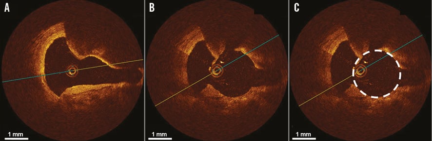
Figure 1. Calcified plaque ablated by rotational atherectomy visualised by optical coherence tomography. A) Optical coherence tomography (OCT) showing lumen narrowing with heavily calcified plaque before rotational atherectomy. After rotational atherectomy, the calcified segment ablated by a 2 mm burr (B) & (C) (dotted line indicates the size of the 2 mm burr).

OCT delineates calcified plaque without acoustic shadowing like intravascular ultrasound. Therefore, OCT is the best intracoronary imaging modality to select the appropriate burr size for RA and also to confirm the efficacy of RA after ablation.

Conflict of interest statement

The authors have no conflicts of interest to declare.

Online data supplement

Moving image 1. Optical coherence tomography after rotational atherectomy clearly visualising ablated calcified plaque.

Supplementary data

To read the full content of this article, please download the PDF.

Optical coherence tomography after rotational atherectomy clearly visualising ablated calcified plaque.

Volume 11 Number 3
Jul 20, 2015
Volume 11 Number 3
View full issue


Key metrics

Suggested by Cory

Debate

10.4244/EIJ-E-24-00014 May 20, 2024
Atherectomy for calcified plaques: orbital for most? Pros and cons
Kirtane A and Ribichini F
free

10.4244/EIJV12I12A237 Dec 20, 2016
Rotational atherectomy: you will never regret using it but you often regret not having used it!
Strisciuglio T and Barbato E
free

IMAGE IN CARDIOLOGY

10.4244/EIJV10I5A104 Sep 28, 2014
High definition OCT image of sharp calcium spicule
Garrido E et al
free
Trending articles
87.2

State-of-the-Art

10.4244/EIJ-D-25-00266 Jan 19, 2026
Lesion stratification with intracoronary imaging
McGarvey M et al
free
47.45

NEW INNOVATION

10.4244/EIJ-D-15-00467 Feb 20, 2018
Design and principle of operation of the HeartMate PHP (percutaneous heart pump)
Van Mieghem NM et al
free
34.8

Original Research

10.4244/EIJ-D-25-01006 Mar 16, 2026
Clinical outcomes and haemodynamic response after blinded stress assessment of moderate aortic stenosis
Eerdekens R et al
22.2

Viewpoint

10.4244/EIJ-D-25-01066 May 4, 2026
Intracoronary imaging guidance for de novo coronary lesion treatment with drug-coated balloons
Amabile N et al
free
20.75

Flashlight

10.4244/EIJ-D-25-01014 Apr 6, 2026
Stent retriever-assisted coronary thrombectomy with continuous aspiration
Liabot Q et al
open access
19.5

Original Research

10.4244/EIJ-D-26-00032 May 15, 2026
Glucocorticoids to reduce permanent pacemaker implantation after TAVI: the GLUCO-TAVI randomised trial
Fuertes-Kenneally L et al
17.8

Expert Review

10.4244/EIJ-D-25-01316 Apr 20, 2026
Electrosurgical laceration and stabilisation of tricuspid edge-to-edge repair: the ELASTA-T technique
Alvarez-Covarrubias H et al
free
X

PCR
Impact factor: 9.5
2024 Journal Citation Reports®
Science Edition (Clarivate Analytics, 2025)
Online ISSN 1969-6213 - Print ISSN 1774-024X
© 2005-2026 Europa Group - All rights reserved