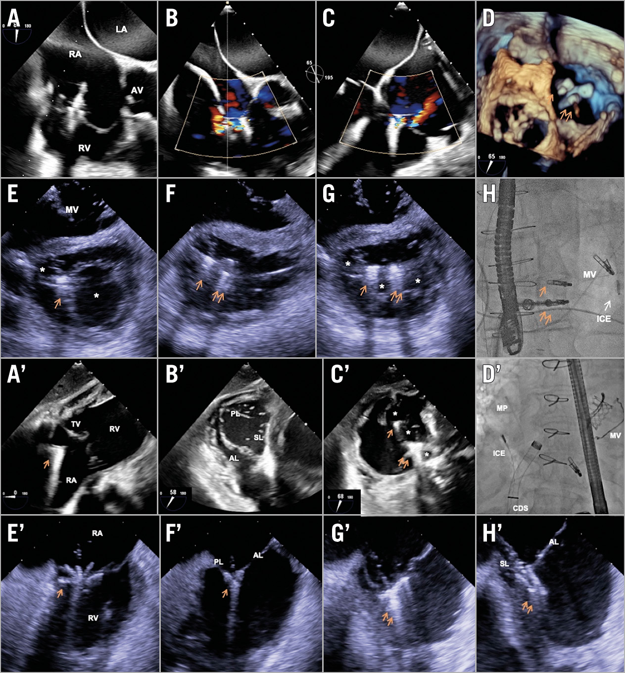
Figure 1. Illustration of the complementary role of ICE in tricuspid edge-to-edge repair. A) – D) Limited TEE transgastric views during a concomitant mitral and tricuspid edge-to-edge repair procedure. E) – H) ICE probe in the LV allowing adequate delineation of the tissue bridge after tricuspid clipping. A’) – D’) Limited TEE mid-oesophageal views during an isolated tricuspid edge-to-edge repair. E’) – H’) ICE imaging from the RA showing excellent grasping views. * new orifice(s). Single arrow indicates first clip. Double arrow indicates second clip. AL: anterior leaflet; AV: aortic valve; CDS: clip delivery system; ICE: intracardiac echo; LA: left atrium; LV: left ventricle; MP: multipurpose catheter; MV: mitral valve; PL: posterior leaflet; RA: right atrium; RV: right ventricle; SL: septal leaflet; TEE: transoesophageal echo; TV: tricuspid valve
Transoesophageal echocardiography (TEE) is the standard imaging modality for procedural guidance of tricuspid edge-to-edge repair. Typically, two TEE views are necessary: a mid-oesophageal view to guide leaflet grasping, and a transgastric view to assess the adequacy of tissue bridging. However, TEE imaging, at least in one of these views, can be challenging due to the remote location of the tricuspid valve from the oesophagus, the degree of right heart enlargement and cardiac rotation, and the acoustic shadowing from cardiac prostheses. In such situations, intracardiac echocardiography (ICE) can provide complementary images to facilitate the procedure1.
Figure 1A-Figure 1D and Moving image 1 illustrate excellent leaflet grasping views from the mid-oesophagus during a concomitant mitral and tricuspid edge-to-edge repair but poor transgastric image quality precluding proper assessment of tissue bridging. In this scenario, a 10 Fr ICE probe (AcuNav™; Siemens, Mountain View, CA, USA) was advanced via the left femoral vein across the iatrogenic transseptal defect (residual from the mitral edge-to-edge repair) into the left ventricle. This allowed visualisation of the tissue bridge and placement of two MitraClip™ XTR (Abbott Vascular, Santa Clara, CA, USA), reducing the tricuspid regurgitation (TR) to mild (Figure 1E-Figure 1H, Moving image 2).
Figure 1A’-Figure 1C’ and Moving image 3 demonstrate excellent transgastric views but suboptimal mid-oesophageal views, limiting the operator’s ability to grasp the leaflets during isolated tricuspid edge-to-edge repair. In this scenario, an ICE probe in the mid right atrium clearly showed grasping of the anterior-posterior leaflets with the clip arms/grippers (Figure 1D’-Figure 1F’). Clockwise rotation of the ICE probe showed grasping of the anterior-septal leaflets (Figure 1G’, Figure 1H’, Moving image 4), facilitating the successful placement of two MitraClip XTR devices.
Conflict of interest statement
The authors have no conflicts of interest to declare.
Supplementary data
To read the full content of this article, please download the PDF.
Moving image 1. Limited TEE transgastric views during tricuspid edge-to-edge repair.
Moving image 2. ICE views showing adequate tissue bridge.
Moving image 3. Limited TEE mid-oesophageal views during tricuspid edge-to-edge repair.
Moving image 4. ICE views showing adequate leaflet grasping.