Coronary angiography and histology form the pillars upon which our present knowledge about coronary atherosclerosis and stenting procedures is based; despite the obvious limitation of both techniques, with angiography being just an imaging modality showing the cast of treated arteries and histology a post mortem assessment that cannot provide in vivo findings.
Based on these assumptions, it is not surprising that one of the most revolutionary innovations in coronary interventions, the use of stents as the most efficient solution to treat a diseased artery, was derived from the application of intravascular ultrasound (IVUS) rather than angiography. Even with a resolution at least ten times less than optical coherence tomography (OCT), IVUS showed that stent thrombosis was due to the poor expansion of stented segments, something that angiography was unable to detect.
OCT opened a new window for the theatre of coronary intervention1, and the four studies collected in the present issue of EuroIntervention represent a clear demonstration of how intriguing it is to navigate inside an artery with the ability to recognise vessel components at an almost cellular level. However, consistent with other studies, this series of interesting papers focused on OCT show that the adoption of this new angle of view generates new doubts, as well as, puzzling researchers with novel images or concepts that have to be interpreted. The search for a more comprehensive understanding of the mechanism of intervention and vessel healing is undoubtedly facilitated by the adoption of high resolution imaging, but unfortunately, the more we see the more the problem become complex. Not all of these OCT images are easy to interpret and classify. The presence of tissue prolapse through the stent struts is an example. As the composition of the tissue cannot be identified with certainty, some authors prefer to avoid the term thrombus prolapse, even in the presence of an intraluminal pedunculated formation. Also, the OCT classification of strut apposition does not have a definitive consensus. In the effort to clarify some OCT images, Radu et al2 have attempted to develop a new analysis of strut apposition with OCT, applying a new methodology to reconstruct stent strut thicknesses and vessel wall areas. Using this method, four types of strut apposition could be identified. Furthermore, the authors have classified a variety of malapposition - including one which has a flower shape pattern as pseudo-apposition. A new underlying mechanism concerning the presence of fibrin tissue separating the stent struts from the original vessel wall was then proposed by this paper.
In the ongoing effort to identify the reasons for drug eluting stent (DES) thrombosis, Kyono et al3 explored the presence of stent coverage in the presence of coronary bifurcations, a well known procedural risk factor for thrombosis. This is an important study because it broadens our current knowledge concerning the mechanism and timing of stent coverage, which inevitably serves as a stimulus to further understand the clinical significance of stent coverage. Here, the authors compared different stent designs: drug eluting stents releasing paclitaxel (PES), sirolimus (SES), zotarolimus (ZES), as well as, bare metal stents (BMS). PES had a greater number of uncovered struts at the ostial site of bifurcations, as compared to other DES and bare stents. They commented on the results with caution, stressing that a clinical link between strut coverage and stent thrombosis had never been proved in prospective study. Yet the correspondence between strut coverage observed in follow-up (FU) OCT studies and other clinical data obtained from prospective clinical trials conducted without OCT is rather clear.
First generation stents have a thrombosis rate much greater than ZES and obviously BMS, with PES having an incidence of late thrombosis slightly higher than SES. Six months FU OCT data shows a percentage of uncovered struts less than 10% after SES, whilst ZES and BMS show complete coverage4. Consistent with these data, similar figures were reported in the REVEAL study (the “Late Breaking Clinical Trial” session, EuroPCR, Barcelona 2009), when the amount of strut coverage was studied at one month follow-up. BMS were found to achieve an earlier coverage than first generation stents. Unfortunately, at the present stage in the evolution of our technology we are not in a position to speculate on the composition of tissue coverage. In their attempt to address tissue composition, the texture analysis that the authors use may not be the right tool to solve the problem. Also, the fact that there is a higher incidence of less coverage at the bifurcation level matches well with the clinical observation that bifurcations represent an anatomical condition with a higher incidence of thrombosis. This paper is, in our view, an additional demonstration that, regardless of the tissue composition, the presence of coverage matters.
Parodi et al5 provide further data on the mechanism and timing of stent coverage, exploring with OCT the left main, a poorly studied portion of the coronary tree that, for technical reasons, the previous occlusive modality was unable to study. The percentage of uncovered struts in the left main was slightly higher than that observed at six months in studies conducted in more distal segments. This is likely due to the 3.5% incidence of malapposition, a feature that is associated to lack of coverage. In the near future, OCT may become a modality to optimise stent positioning due to the recent introduction of the frequency domain technique. It is obvious that in order to do so, OCT must prove to be capable of addressing also the left main. It is for this reason we should congratulate the authors, who were able to study the left main using time domain OCT, with all the limitations inherent in this previous generation technique. It has to be emphasised that the authors report on one of the major limitation of time domain OCT – the drop out of vessel contour in the presence of a large vessel and/or eccentricity of the source – which has been overcome by the novel frequency domain technology. The only problem (not trivial) we still see for left main OCT assessment is the impossibility of studying lesions very close to the ostium, which, at the current stage of the technology – and even with frequency domain OCT – remain almost impossible to study.
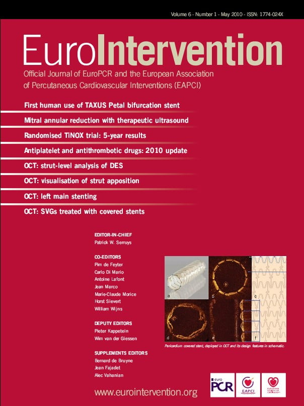
Tyczynski et al6 contribute to the concept that the use of OCT can definitely help in understanding the mechanism of coronary interventions. The use of pericardium covered stents is a reasonable solution in the presence of degenerated saphenous grafts. The authors show that luminal defects observed after pericardium covered stents were due to a bulging of the pericardium between the struts, with the soft atherosclerotic tissue being trapped between the pericardial layer and the saphenous wall. OCT was able to exclude thrombi as the reason of luminal narrowing after stenting and this, apart the contribution to our understanding of the mechanism of how a new stent designs works, shows the potential of OCT in a OCT guided approach.
Therefore the manuscript indirectly raises a question: Can we use the burden of information provided by OCT to improve interventions? And if yes, in which clinical and anatomical scenarios? It is possible that the excellent superficial definitions of coronary arteries provided by OCT will become a valid approach to improving interventions by providing accurate luminal measurements as well as addressing details that may be missed by IVUS, such as plaque or thrombus prolapse inside the stent or at the margins, or dissections. Preliminary data from our group, obtained in 120 patients treated with OCT guided procedures, showed the approach to be safe and feasible, with 28% of cases requiring further intervention (balloon dilatation or stent deployment), because – despite the achievement of an optimal angiographic result – OCT revealed an insufficient result with underexpansion, edge dissection or marked plaque prolapse.
There is obviously a need for more data to support an OCT-guided approach to stenting. OCT criteria must be validated, and randomised studies where OCT is compared to quantitative coronary angiography or IVUS will verify whether this is a valid approach7.