
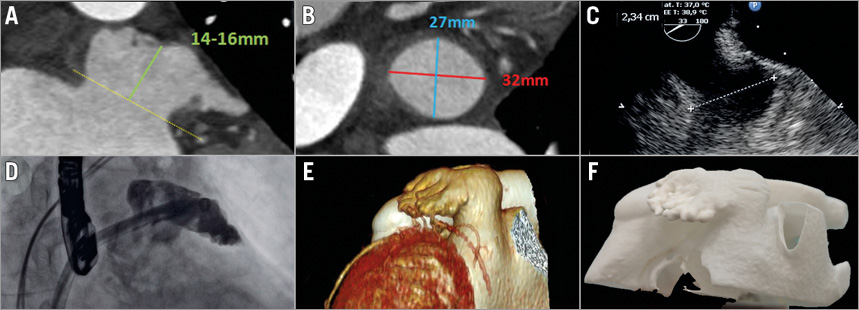
An 84-year-old female with paroxysmal atrial fibrillation, CHA2DS2-VASc score 4, HAS-BLED score 3, developed subdural haematoma on warfarin and was qualified for left atrial appendage occlusion (LAAO). The first attempt at LAAO was abandoned due to complex appendage anatomy with a large-sized landing zone and chicken-wing shape. This prompted obtaining a 3D-printed polyamide model of the heart (Panel A-Panel F). Rehearsals were undertaken in the model with 31 mm and 34 mm ACP Amulet™ devices (St. Jude Medical, St. Paul, MN, USA) (Online Figure 1). Contrary to the sizing chart, assessment of the devices’ position in the model indicated the 31 mm device as optimal, while a stable position could not be achieved with the 34 mm device. A 31 mm device was delivered to the patient but a leak was noted (Online Figure 2A, Online Figure 2B). Experience with the 3D model guided operators on device redeployment with counterclockwise rotation of the sheath (Online Figure 2C, Online Figure 2D) and complete appendage sealing (Online Figure 3).
Acknowledgements
St. Jude Medical provided sheaths and left atrial appendage occluders for rehearsals in the 3D-printed model free of charge.
Conflict of interest statement
R. Pracon has received speaker’s honoraria from St. Jude Medical. M. Demkow is a proctor for St. Jude Medical. The other authors have no conflicts of interest to declare.
Supplementary data
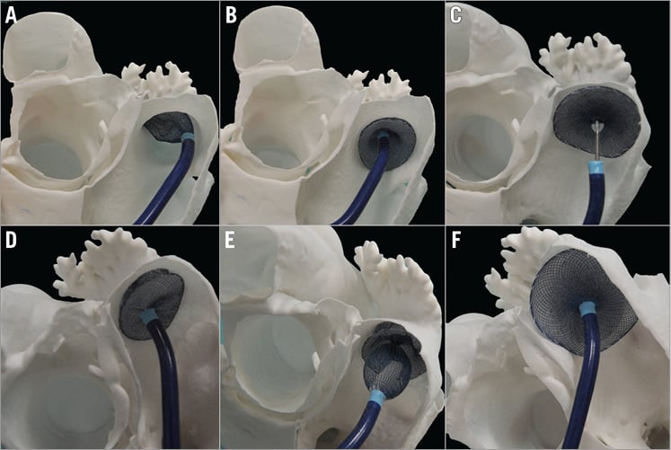
Online Figure 1. Attempts to occlude the appendage in the 3D-printed model. A) - C) 31 mm, well-fitted device. D) - F) 34 mm, oversized device.
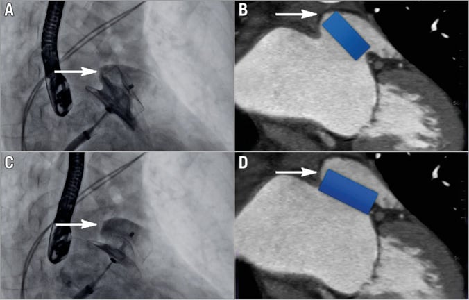
Online Figure 2. Intraprocedural imaging and computed tomography with superimposed, draft device position. A) & B) After the first deployment (arrow indicates suboptimal position of superior device’s edge and a leak). C) & D) After device redeployment (arrow indicates optimal device position without the leak).
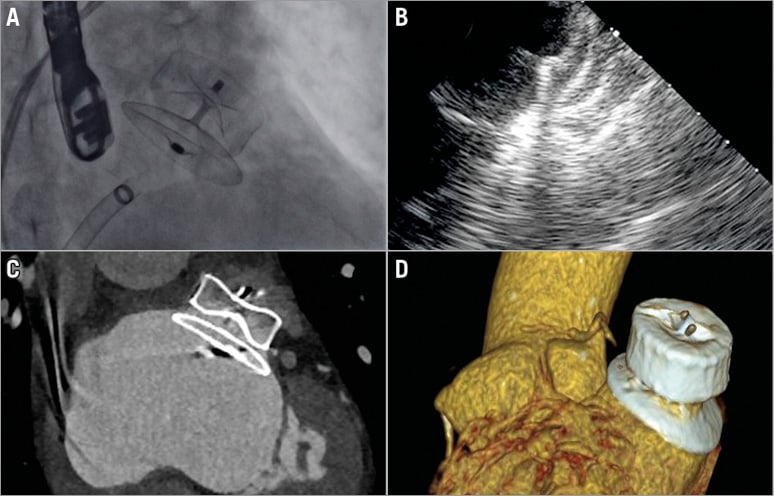
Online Figure 3. Post-procedural imaging. A) Fluoroscopy. B) Transoesophageal echocardiography. C) Computed tomography (CT). D) 3D CT reconstruction.
Moving image 1. Angiography of the left atrial appendage.
Moving image 2. First deployment of the 31 mm ACP Amulet device’s lobe.
Moving image 3. First deployment of the 31 mm ACP Amulet device’s disc.
Moving image 4. Angiography after the first deployment of the 31 mm ACP Amulet device in the left atrial appendage with visible leak over the superior portion of the device.
Moving image 5. Counterclockwise rotation of the sheath and second deployment of the 31 mm ACP Amulet device’s lobe.
Moving image 6. Second deployment of the 31 mm ACP Amulet device’s disc.
Moving image 7. Final angiography after the second deployment of the 31 mm ACP Amulet device in the appendage; no visible leak over the superior portion of the device.
Moving image 8. The 31 mm ACP Amulet device in the appendage after its release from the delivery cable.
Supplementary data
To read the full content of this article, please download the PDF.
Angiography of the left atrial appendage.
First deployment of the 31 mm ACP Amulet device’s lobe.
First deployment of the 31 mm ACP Amulet device’s disc.
Angiography after the first deployment of the 31 mm ACP Amulet device in the left atrial appendage with visible leak over the superior portion of the device.
Counterclockwise rotation of the sheath and second deployment of the 31 mm ACP Amulet device’s lobe.
Second deployment of the 31 mm ACP Amulet device’s disc.
Final angiography after the second deployment of the 31 mm ACP Amulet device in the appendage; no visible leak over the superior portion of the device.
The 31 mm ACP Amulet device in the appendage after its release from the delivery cable.