IMAGE IN CARDIOLOGY

DOI: 10.4244/EIJ-D-16-00263

Coronary magnetic resonance angiographic images of a lesion treated with a metal stent and fully bioresorbable polymeric everolimus-eluting scaffold

Jiro Aoki1*, MD, PhD; Satoru Kishi2, MD; Shigeru Saito3, MD, PhD; Kengo Tanabe1, MD, PhD

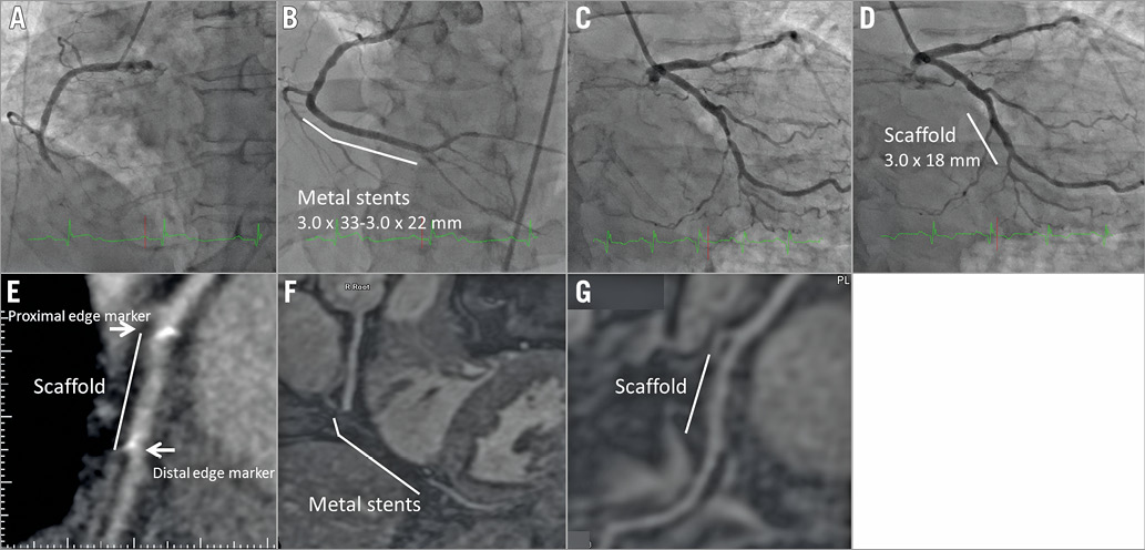
A 48-year-old man was referred to our hospital for acute coronary syndrome. Coronary metal stents were implanted at the distal right coronary artery as the culprit lesion (pre-treatment: Panel A; post-treatment: Panel B). After one month, he was readmitted to our hospital in order to treat the remaining coronary artery stenosis. He was enrolled in the ABSORB EXTEND study, and a fully bioresorbable polymeric everolimus-eluting scaffold (Absorb; Abbott Vascular, Santa Clara, CA, USA) was implanted at the mid left circumflex coronary artery (pre-treatment: Panel C; post-treatment: Panel D). Follow-up coronary computed tomographic angiography was performed at eight months after the scaffold implantation. Mild stenosis and small platinum markers at scaffold edges were depicted at the mid left circumflex coronary artery (Panel E). At three years following the scaffold implantation, coronary magnetic resonance angiography (MRA) was performed to check his coronary artery. A coronary luminal image was not obtained at the metal stent site due to metal artefact (Panel F). However, a coronary luminal image was clearly obtained at the scaffold site (Panel G). No restenosis was observed, and the small platinum markers did not hamper lumen visibility. The patient is currently doing well without symptoms. Coronary MRA does not require radiation exposure and a contrast agent. Coronary MRA might be a useful modality to follow patients treated with fully bioresorbable polymeric scaffolds.

Conflict of interest statement

K. Tanabe is a member of the Advisory Board of Abbott Vascular Japan and receives lecture honoraria from Abbott Vascular Japan. The other authors have no conflicts of interest to declare.

Volume 12 Number 12
Dec 20, 2016
Volume 12 Number 12
View full issue


Key metrics

On the same subject

10.4244/EIJV11I12A259 Mar 18, 2016
Oversized post-dilatation of current bioresorbable vascular scaffolds: kill or cure?
von Birgelen C and Basalus M
free

Editorial

10.4244/EIJ-E-23-00025 Jun 19, 2023
Novel bioresorbable scaffolds and lessons from recent history
Joner M et al
free
Trending articles
338.63

State-of-the-Art Review

10.4244/EIJ-D-21-00904 Apr 1, 2022
Antiplatelet therapy after percutaneous coronary intervention
Angiolillo D et al
free
295.45

Expert consensus

10.4244/EIJ-D-21-00898 Sep 20, 2022
Intravascular ultrasound guidance for lower extremity arterial and venous interventions
Secemsky E et al
free
226.03

State-of-the-Art Review

10.4244/EIJ-D-21-00426 Dec 3, 2021
Myocardial infarction with non-obstructive coronary artery disease
Lindahl B et al
free
209.5

State-of-the-Art Review

10.4244/EIJ-D-21-01034 Jun 3, 2022
Management of in-stent restenosis
Alfonso F et al
free
168.4

Expert review

10.4244/EIJ-D-21-00690 May 15, 2022
Crush techniques for percutaneous coronary intervention of bifurcation lesions
Moroni F et al
free
150.28

State-of-the-Art

10.4244/EIJ-D-22-00776 Apr 3, 2023
Computed tomographic angiography in coronary artery disease
Serruys PW et al
free
103.48

Expert consensus

10.4244/EIJ-E-22-00018 Dec 4, 2023
Definitions and Standardized Endpoints for Treatment of Coronary Bifurcations
Lunardi M et al
free
X

The Official Journal of EuroPCR and the European Association of Percutaneous Cardiovascular Interventions (EAPCI)

EuroPCR EAPCI
PCR ESC
Impact factor: 6.2
2022 Journal Citation Reports®
Science Edition (Clarivate Analytics, 2023)
Online ISSN 1969-6213 - Print ISSN 1774-024X
© 2005-2024 Europa Group - All rights reserved