Introduction
Alternative approaches for mitral valve repair are constantly being developed. Even though the safety of these devices has been extensively assessed in healthy animals before approval for first-in-man studies, often the efficacy of these new devices has hardly been challenged in vivo in the setting of mitral regurgitation (MR). Therefore, reliable and reproducible preclinical animal models of MR are crucial to allow the safety assessment and efficacy evaluation of these novel approaches.
Material and methods
Eight young female Landrace domestic pigs (60±12 kg) were used. A control group with age, sex and weight-matched animals was constituted after the six-week follow-up period (n=7). The experimental protocol was approved by the local authority ethics committee.
To obtain an ischaemic MR, a localised posteromedial papillary muscle (PMPM) myocardial infarction (MI) involving the underlying inferior wall was achieved. Contrast echocardiography was used as a PMPM vascularisation mapping tool. The obtuse marginal (OM) branches irrigating the PMPM were identified by selectively injecting regular radiographic contrast agent through an over-the-wire (OTW) balloon. Echocardiography allowed detection of any contrast enhancement of the area of interest. To achieve MI, 1 to 2 ml of pure ethanol was slowly injected into each target branch through the OTW balloon after inflation. The balloon was left in place for 15 minutes, then deflated (Figure 1).
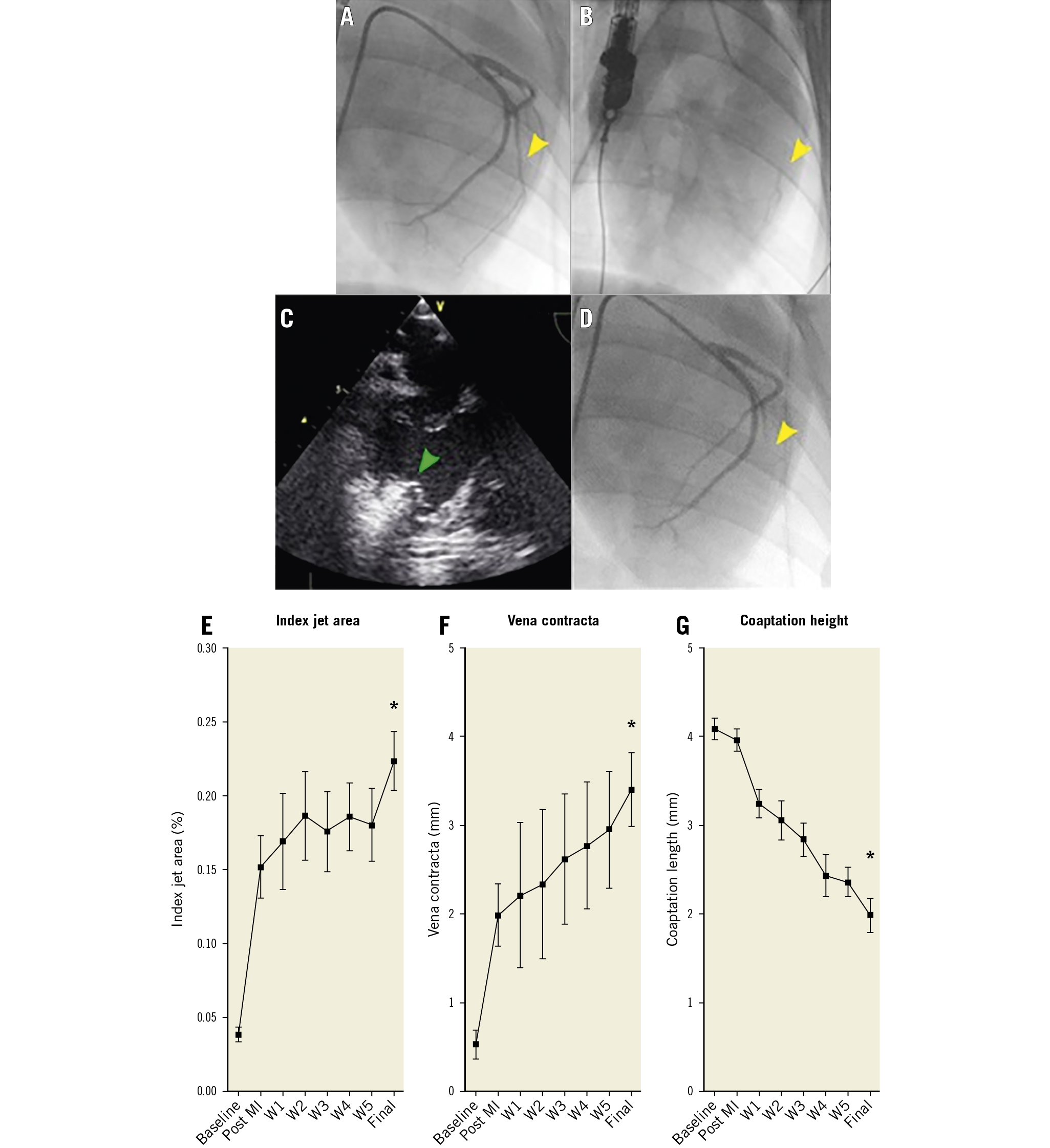
Figure 1. Ischaemic MR induction procedure supported by contrast echocardiography and its evolution. Representative baseline LCA angiogram (A). Injection of contrast media in the obtuse marginal (OM) branch (yellow arrow) through an OTW balloon (B) and corresponding contrast enhancement on the echocardiography (green arrow) (C). Result after injection of ethanol (D). Temporal evolution of index jet area (E), vena contracta (F) and coaptation height (G). * p<0.001 ANOVA.
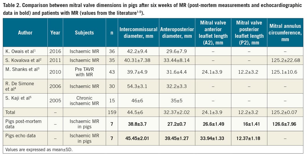
Six weeks post MI, the animals were sacrificed and the hearts harvested. Mitral valve diameters were measured and compared to data from human patients from the literature [1–5].
Results
All the animals survived the acute phase. One animal was euthanised at two weeks after an acute pulmonary oedema.
Table 1 summarises the echocardiographic findings. A semiquantitative method was used to evaluate and monitor MR severity and evolution. A moderate ischaemic MR was obtained at the end of the follow-up period. Parallel to MR development, valve tethering evolved progressively as reflected by the significantly increased tenting height and area concordant with an ischaemic MR (Figure 2).
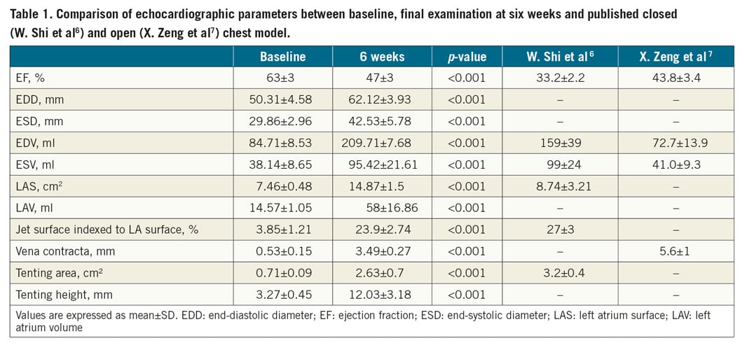
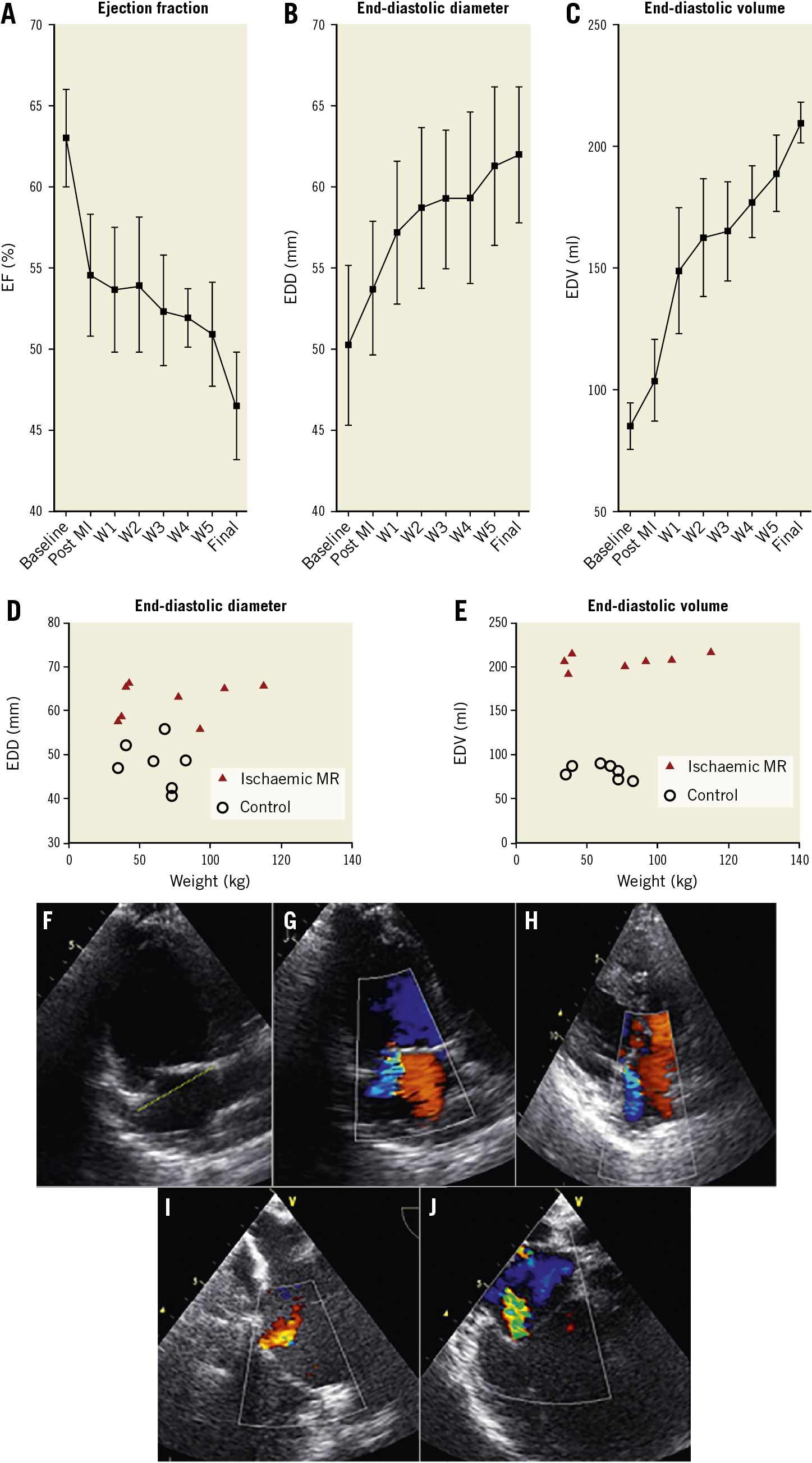
Figure 2. Echocardiographic follow-up of ischaemic MR and LV function and dimensions. Temporal evolution of ejection fraction (A), LV end-diastolic diameter (B) and volume (C). Distribution of LV end-diastolic diameter (D) and volume (E) in mitral regurgitation pigs (red triangles) and age, sex and weight-matched animals (empty circles). Representative echocardiograms of the tenting below the annular plane (yellow dotted line) (F). Representative TTE colour Doppler of MR after six weeks (G & H) and TEE (I & J).
As expected, the echo values of the mitral annulus diameter were slightly higher than the ones obtained post mortem and showed a range of values close to the human data (Table 2). Post-mortem measurement of the anterior leaflet in the pigs showed similar lengths to those of human patients, as opposed to the posterior leaflet which was found to have a longer length in pigs.
Discussion (Supplementary Appendix 1)
We established for the first time a closed chest pig model of ischaemic MR guided by contrast echocardiography. Echo mapping of the PMPM vasculature allowed achieving a controlled and reproducible MI of the PMPM with consistent development of a significant MR in all animals.
Our model did not rely exclusively on fluoroscopic guidance while identifying the irrigating branches as in the previous ethanol injection model described by W. Shi et al6. Intraoperative contrast echocardiography support allowed more reliable identification of target branches.
Pig hearts are widely accepted for preclinical studies given their close similarities to human hearts. In our model, mitral annular diameter values were in a range close to the values measured in MR patients. Therefore, this model can easily accommodate mitral devices designed for humans.
Limitations
Our study was limited by the growth rate of the animals and resulted in a short follow-up period which did not allow the development of a higher MR grade. A longer follow-up period might be necessary to observe a more severe MR.
Conclusion
We established a novel ischaemic MR model in pigs using contrast echocardiography guidance. This model could be a suitable platform for testing new mitral valve devices.
|
Impact on daily practice Transcatheter alternatives for ischaemic MR are the optimal solution for surgical high-risk patients. We provide a reproducible model of ischaemic MR which can serve as a relevant platform to test transcatheter therapy efficacy before first-in-man trials. |
Funding
The study was supported by a grant from the Ludwig Boltzmann Society (REM2017-20).
Conflict of interest statement
The authors have no conflicts of interest to declare.
Supplementary data
To read the full content of this article, please download the PDF.