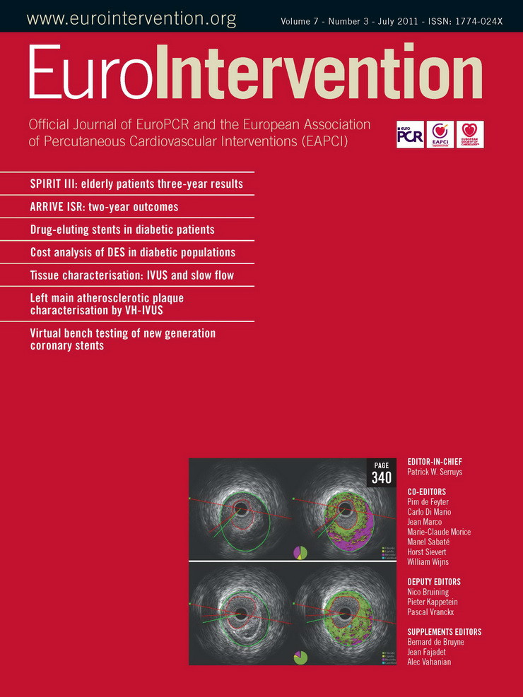
Intravascular ultrasound (IVUS) imaging permits the assessment of coronary atherosclerosis in vivo1, and for nearly two decades greyscale IVUS has been used to guide percutaneous coronary interventions (PCI), especially in challenging lesion subsets such as major bifurcations2. More recently, spectral analysis of radiofrequency IVUS data (RF-IVUS) has been used to obtain further quantitative information on coronary plaque components (e.g., necrotic core), which has permitted characterisation of certain plaque phenotypes such as the “vulnerable” thin-cap fibroatheroma (TCFA)1,3,4. The left main (LM) stem has extensively been studied with IVUS1,5-16. The reasons for that are the significant impact of LM disease on morbidity and mortality, and the known diagnostic difficulty of coronary angiography in assessing intermediate LM disease14-16.
Cardiovascular event risk and mortality increase markedly when a LM stenosis reaches haemodynamic significance16. The challenge of determining the significance of an angiographically intermediate LM stenosis has been substantially improved by the use of IVUS imaging and pressure wire-based measurements of fractional flow reserve (FFR)14-17. Based on previous IVUS studies in LM lesions, a minimum lumen cross-sectional area of less than 6 mm² was considered the threshold to identify lesions that require revascularisation. However, preliminary data presented at EuroPCR 2011 question this IVUS threshold and suggest that measurement of FFR is superior18. IVUS, on the other hand, provides useful information on lesion characteristics, such as the amount and distribution of lesion calcium, and the involvement of side branches and total vessel size. Such information can guide or alter PCI strategies, especially in LM bifurcation lesions2. As a consequence, many interventional cardiologists currently determine the need for LM revascularisation based on FFR measurements, while IVUS is used to guide LM PCI2,17,18. Today, optical coherence tomography (OCT) is considered an alternative to IVUS for the assessment of the result of stenting. However, in large and proximally located coronary segments, such as the LM stem or the ostium of the right coronary artery, IVUS implies evident advantages over OCT19.
Yet what is the fate of mild and FFR-negative LM lesions? Previous serial studies with conventional greyscale IVUS provided insight into the natural course of such mild LM plaques1,5,7-9. For instance, serial observational IVUS data suggested a direct linear relationship between low density lipoprotein-cholesterol levels and the progression of mild LM plaques5, which was subsequently confirmed by randomised clinical trials1. Imaging with IVUS also permits the assessment of compensatory vascular remodelling, which is considered an important feature of both vulnerable lesions and ruptured coronary plaques8-13,20-22. A broad spectrum of true vascular remodelling was demonstrated with serial IVUS; in particular, lumen size of mildly diseased LM coronary arteries depended more on the direction of vessel remodelling than on plaque progression8,9,13. Interestingly, the direction of (serial) remodelling, i.e., compensatory vascular enlargement versus vessel shrinkage, could not be predicted by the quantity of baseline plaque burden9,21. In an IVUS study by Ricciardi et al6, angiographically silent LM atherosclerosis was an independent predictor of future adverse events. But which features of such non-significant LM plaques may affect outcome or could be considered a surrogate marker candidate of future adverse events? Hong et al10 found that a non-negative remodelling state was a predictor of future LM-related coronary events. In addition, our group observed a significant relation between the progression of mild LM plaques, as measured by serial IVUS, and various cardiovascular risk scores7.
However, regardless of progression rate and geometrical lesion characteristics, the presence of certain plaque components and/or plaque phenotypes may augment or reduce the coronary risk. Use of RF-IVUS allows us to assess and quantify different plaque components in vivo and to identify plaque phenotypes that could be precursors of coronary events1,3,4. In the current edition of the journal, Mercado et al23 used RF-IVUS to obtain interesting insights into plaque composition and the presence of certain “high-risk” plaque phenotypes in LM stems and adjacent proximal left anterior descending (LAD) coronary segments. The authors report that TCFA and necrotic core content, which are both considered surrogate markers of plaque vulnerability, were located predominantly in the proximal LAD rather than in the LM. These findings underline previous histopathological and clinical observations, suggesting that the proximal LAD is a predilection site of plaque rupture24. In addition, the data of Mercado et al23 are in good agreement with findings of Valgimigli et al11, who also observed more necrotic core content in proximal LAD segments.
But what is the predictive value of demonstrating such TCFA in haemodynamically non-significant lesions? The PROSPECT trial recently demonstrated an association between RF-IVUS derived TCFA (in non-culprit lesions) and adverse events25. This finding suggests that RF-IVUS findings can have a certain predictive value, but there is still no indication for invasive treatment of haemodynamically non-significant coronary lesions with TCFA phenotype. In fact, observational non-serial RF-IVUS data (from a single point in time) do not depict the dynamic nature of the disease. For instance, the amount of necrotic core at baseline does not represent necrotic core expansion over time, and a potential transition to more “vulnerable” plaque phenotypes can only be identified during serial examinations. A recent serial RF-IVUS study by Kubo et al26 in non-obstructive non-culprit lesions underlines this limitation, as the majority of TCFA showed “healing” during follow-up and transformed to “less vulnerable” plaque phenotypes, while some “less vulnerable” stages of atherosclerotic disease transformed into TCFA .
In significant coronary lesions prior to PCI, however, it may be particularly interesting to obtain information on plaque composition. Such data could identify lesions at increased risk of PCI-related complications, which may help to tailor interventional procedures27,28. In the current issue of the journal, Utsunomiya et al28 report interesting RF-IVUS data of patients with acute coronary syndrome, showing a relation between the necrotic core volume of culprit lesions before PCI and the occurrence of no-reflow during PCI. PCI-related complications such as the no-reflow phenomenon are particularly feared in the setting of LM PCI, as they may affect the circulation of the entire left coronary system. While in clinical practice the frequency of LM PCI is increasing, risk stratification of such interventions may be particularly important. Angiography-based risk stratification of multivessel PCI, such as the use of the SYNTAX score29, was shown to be valuable and is currently being studied in the setting of LM PCI (EXCEL trial)30. Future prospective studies may address the question of whether the use of RF-IVUS based information on plaque composition may help to improve risk stratification of PCI in major coronary segments such as the left main stem.
Conflict of interest statement
The authors have no conflict of interest to declare.
References

