Editorial

DOI: 10.4244/EIJ-E-25-00045

Angiographic-based assessment of microvascular dysfunction: quo vadis?

Christos V. Bourantas1,2, MD, PhD; Yoshinobu Onuma3,4, MD, PhD; Hector M. Garcia-Garcia5, MD, PhD; Patrick W. Serruys3,4, MD, PhD

Over recent years there has been a revolution in the applications of coronary angiography in the assessment of coronary artery pathology. Computerised methodologies have been introduced that are able to process coronary artery models reconstructed from angiographic images and accurately evaluate lesion haemodynamic severity, plaque deformability, and peripheral vascular resistance. Despite the inherent limitations of angiography to quantify lumen dimensions and measure coronary flow, cumulative evidence supports the validity of this concept. Methodologies developed to compute the fractional flow reserve (FFR) are now indicated by European guidelines in treatment planning: radial wall strain, which measures the changes in lumen dimensions during the cardiac cycle in order to assess the mechanical properties of plaque, has been found able to detect vulnerable lesions1, while solutions that were proposed to measure microvascular resistance from coronary angiography (Figure 1) seem able to provide useful prognostic information and predict microvascular obstruction (MVO) and infarct size post-revascularisation in patients with an acute coronary syndrome234.

In this issue of EuroIntervention, Zhang et al examined for the first time the efficacy of the...

Sign in to read
the full article

Forgot your password?
No account yet?
Sign up for free!

Create my pcr account

Join us for free and access thousands of articles from EuroIntervention, as well as presentations, videos, cases from PCRonline.com
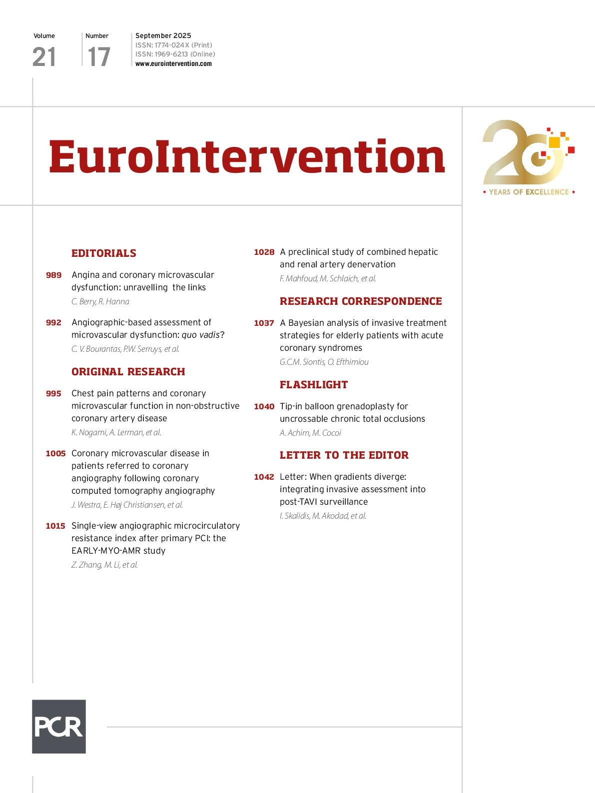
Volume 21 Number 17
Sep 1, 2025
Volume 21 Number 17
View full issue


Key metrics

Suggested by Cory

EXPERT REVIEW

10.4244/EIJ-D-17-00465 Aug 25, 2017
State of the art: coronary angiography
Collet C et al
free

EXPERT REVIEW

10.4244/EIJ-D-17-00503 Aug 25, 2017
State of the art: pressure wire and coronary functional assessment
Echavarria-Pinto M et al
free

Clinical Research

10.4244/EIJ-D-21-00471 Apr 22, 2022
Vessel fractional flow reserve (vFFR) for the assessment of stenosis severity: the FAST II study
Masdjedi K et al
free

10.4244/EIJV13I2A20 Jun 2, 2017
Coronary physiological parameters at a crossroads
Davies J et al
free
Trending articles
318.3

State-of-the-Art Review

10.4244/EIJ-D-21-00695 Nov 19, 2021
Transcatheter treatment for tricuspid valve disease
Praz F et al
free
116.85

State-of-the-Art

10.4244/EIJ-D-24-00066 Apr 21, 2025
Management of complications after valvular interventions
Bansal A et al
free
108.3

Viewpoint

10.4244/EIJ-E-22-00007 May 15, 2022
TAVI at 20: how a crazy idea led to a clinical revolution
Eltchaninoff H et al
free
91.6

Image – Interventional flashlight

10.4244/EIJ-D-22-00344 Aug 5, 2022
First dedicated transcatheter leaflet splitting device: the ShortCut device
Tchétché D et al
free
72.15

State-of-the-art

10.4244/EIJ-D-22-00627 Feb 6, 2023
Left atrial appendage occlusion
Holmes D et al
free
68.9

State-of-the-Art

10.4244/EIJ-D-24-00992 Sep 15, 2025
Antithrombotic therapy in complex percutaneous coronary intervention
Castiello D et al
free
49.55

CLINICAL RESEARCH

10.4244/EIJ-D-17-00962 Apr 6, 2018
A new optical coherence tomography-based calcium scoring system to predict stent underexpansion
Fujino A et al
free
42.95

State-of-the-Art Review

10.4244/EIJ-D-21-00145 Sep 20, 2021
Robotics, imaging, and artificial intelligence in the catheterisation laboratory
Beyar R et al
free
X

PCR
Impact factor: 9.5
2024 Journal Citation Reports®
Science Edition (Clarivate Analytics, 2025)
Online ISSN 1969-6213 - Print ISSN 1774-024X
© 2005-2025 Europa Group - All rights reserved