Percutaneous left atrial appendage (LAA) occlusion (LAAO) is an alternative to oral anticoagulation for those patients with atrial fibrillation and a contraindication to or high risk for anticoagulation1. LAAO procedures are typically performed with a 3D imaging technique (transoesophageal echocardiography [TOE] or computed tomography [CT]). For intraprocedural guidance, 3D-TOE is generally used, requiring general anaesthesia in most centres. Technological improvements such as the micro-TOE probe (10T-D; GE Healthcare)2, allow LAAO guidance using only minimal sedation and resulting in same-day patient hospital discharge. However, micro-TOE also has limitations (monoplane imaging, bad visualisation of the far field, need for specialised training, etc.) which limit intraprocedural guidance in complex anatomies or inexperienced centres.
A novel mini TOE probe with 3D capabilities (9VT-D; GE Healthcare) has recently been released on the market for use in the paediatric population (Figure 1A) but with the potential to be used in adults as well. Theoretically, it will maintain the good tolerance of the micro-TOE probe while providing full 3D imaging capabilities. There are additional advantages, as compared to the micro-TOE probe, that include superior 2D imaging quality, bi-plane imaging for a safer transseptal puncture and better control during device implantation, and the possibility to perform 3D measurements for device selection during the intervention.
We present a series of the first 4 patients who underwent LAAO guided with a 3D mini TOE probe at our centre. All patients were consecutively treated on the same day. The tolerance of the probe was excellent for all of the patients, with local oropharyngeal anaesthesia with lidocaine and minimal sedation (0.025-0.05 mg of fentanyl and 1-2 mg of midazolam − the same regimen used with the micro-TOE probe at our centre). Imaging was good, allowing a safe transseptal puncture with bi-plane guidance and 3D measurement of the landing zone. Device sizing was based on the 3D measurements obtained during the intervention. All four procedures were successful with no residual leaks and no intraprocedural complications (mean duration of the intervention 52 min, fluoroscopy 12.5 min, radiation dose 58.1 microGy/m2, TOE probe insertion 25.2 min). Only one patient (#2) required angiographic contrast administration due to the extreme chicken wing morphology of the LAA. Figure 1B-Figure 1G and Moving image 1 show the interventional echocardiographic images during LAAO of patient #1. All patients were discharged on the same day with no complications after a 6-hr monitorisation and check-up echocardiogram for pericardial effusion evaluation. Supplementary Table 1 includes patient characteristics and procedural data for all patients; Supplementary Figure 1 includes images from the interventions in patients #2-4.
The initial experience with the new 3D mini TOE probe showed good tolerance (despite minimal sedation and the supine position of the patient) with excellent imaging quality of the LAA that allowed an effective and safe LAAO guidance. The use of mini/micro TOE probes may improve the efficiency of the catheterisation laboratory by increasing the number of patients treated per workday and allowing a safe same-day hospital discharge.
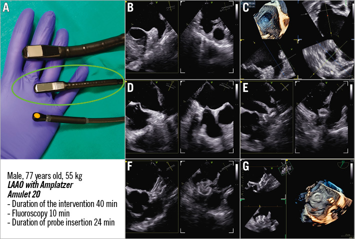
Figure 1. Interventional echocardiographic images during left atrial appendage occlusion of the first patient. A) A comparison of the mini 4D probe 9VT-D (centre, green circle) with the standard 4D probe 6VT-D (top) and the micro 2D probe 10T-D (bottom). TOE images of patient #1 during LAAO: B) LAA with bi-plane view, C) 3D LAA image with multiplanar reconstruction for landing zone measurement, D) bi-plane view for transseptal puncture with the needle inducing tenting in the septum, E) initial positioning of the device with bi-plane view, F) final position of the device with bi-plane and 3D imaging (G). 3D: three-dimensional; LAAO: left atrial appendage occlusion; TOE : transoesophageal echocardiography
Conflict of interest statement
X. Freixa is a proctor for Abbott and LifeTech Science. M. Sitges is a consultant and speaker for General Electric, Medtronic, Edwards Lifesciences, and Abbott. L. Sanchis is a proctor for Abbott and a speaker for General Electric and Abbott. The other authors have no conflicts of interest to declare regarding the present paper.
Supplementary data
To read the full content of this article, please download the PDF.
Moving image 1. The interventional echocardiographic images during LAAO of patient #1.