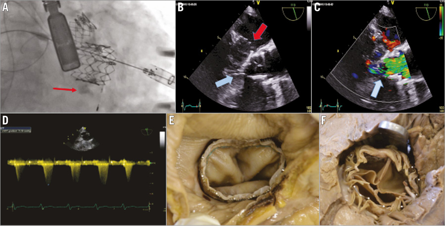

We describe the first case of transcatheter replacement of both aortic and mitral native valves in a 91-year-old female patient with severe symptomatic aortic stenosis and severe mitral stenosis. A CT scan confirmed circumferential mitral annular calcification (MAC). Via a transapical approach, a 23 mm SAPIEN XT valve (Edwards Lifesciences, Irvine, CA, USA) was successfully placed (Moving image 1). The mitral annulus measured 19×26 mm and a 29 mm SAPIEN XT valve was deployed within the MAC (arrow in Panel A, Moving image 2). Transoesophageal echo (TEE) confirmed good function of both valves, but the prosthetic mitral valve (red arrow) caused obstruction of the aortic valve inflow (blue arrow in Panel B, Moving image 3). The same view with colour shows turbulent flow in the left ventricular outflow tract (LVOT) due to the protrusion of the crown of the mitral prosthesis (Panel C, Moving image 4). A gradient of 73.59 mmHg was noted over the LVOT in the TEE transgastric long-axis view (Panel D). The patient was extubated on the table but her blood pressures remained extremely labile and she died 12 hours later. Post-mortem evaluation confirmed good placement of both valves. The mitral valve was compressed by cardiopulmonary resuscitation (CPR) when viewed from the atrial side (Panel E), and in Panel F (from the ventricular side) there is a scalpel handle protruding through the aortic valve into the ventricle showing obstruction of the LVOT.
Conflict of interest statement
The authors have no conflicts of interest to declare.
Supplementary data
Moving image 1. Deployment of the transcatheter aortic valve via a transapical approach.
Moving image 2. Deployment of a reverse-mounted 29 mm SAPIEN XT valve in the clearly visible ring of mitral annular calcification.
Moving image 3. 2D transoesophageal echo (TEE) long-axis view showing the mitral prosthesis functioning well but causing obstruction to the aortic outflow tract.
Moving image 4. The same view as Moving image 3 but with colour flow indicating the acceleration of flow where the crown of the mitral valve obstructs the left ventricular outflow tract.
Supplementary data
To read the full content of this article, please download the PDF.
Deployment of the transcatheter aortic valve via a transapical approach.
Deployment of a reverse-mounted 29 mm SAPIEN XT valve in the clearly visible ring of mitral annular calcification.
2D transoesophageal echo (TEE) long-axis view showing the mitral prosthesis functioning well but causing obstruction to the aortic outflow tract.
The same view as but with colour flow indicating the acceleration of flow where the crown of the mitral valve obstructs the left ventricular outflow tract.

