IMAGE IN CARDIOLOGY

DOI: 10.4244/EIJ-D-16-00067

Intravascular ultrasound and optical coherence tomography images for direct right coronary artery injury after tricuspid valve annuloplasty

Junsuke Shibuya1, MD; Nobuaki Kobayashi1*, MD, PhD; Noritake Hata1, MD, PhD; Shunsuke Uetake2, MD, PhD; Masahiro Fujii3, MD, PhD; Ryuzo Bessho3, MD, PhD; Wataru Shimizu4, MD, PhD

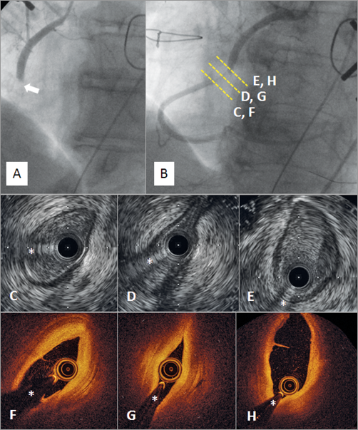
A 76-year-old female was admitted with dyspnoea and leg oedema. Echocardiography showed severe mitral and tricuspid valve regurgitation with marked right ventricular (RV) dilatation, and coronary angiography showed intact left and right coronary arteries (RCA) (Online Figure 1). She was diagnosed with heart failure due to combined valvular disease and underwent tricuspid valve (the Kay bicuspidisation technique without ring) and mitral valve annuloplasty. After the operation, she suffered from myocardial infarction with ST-segment elevation in the inferior leads. Coronary angiography showed a total occlusion (arrow) at the middle portion of the RCA (Panel A, Moving image 1). After passing a wire through the lesion, angiography revealed a bent RCA with tight stenosis (Panel B, Moving image 2). Intravascular ultrasound (Panel C-Panel E, Moving image 3) and optical coherence tomography (Panel F-Panel H, Moving image 4) showed a distorted and collapsed vessel without obstructive thrombi at the bent portion of the RCA. Stenting (Nobori™ 3.5/24 mm; Terumo, Tokyo, Japan) improved coronary flow, with two bends at the middle and distal parts of the RCA (Online Figure 2, Moving image 5). The stent patency was confirmed by coronary angiography five months after the procedure.

Intracoronary images described the distorted and collapsed RCA at the bent part, which was caused by Kay bicuspidisation annuloplasty, rectified by plicating the annulus along the posterior leaflet.

Conflict of interest statement

The authors have no conflicts of interest to declare.

Supplementary data

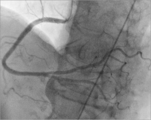
Online Figure 1. Right coronary artery before tricuspid valve annuloplasty.

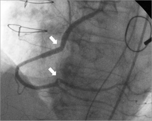
Online Figure 2. Right coronary artery after stenting.

Moving image 1. Angiography of the totally occluded RCA.

Moving image 2. Angiography of wire crossing showing the bent RCA with severe stenosis.

Moving image 3. IVUS showing a distorted and collapsed RCA at the bent area.

Moving image 4. OCT showing a distorted and collapsed RCA at the bent area.

Moving image 5. Angiography after stenting showing improved flow with two bends in the RCA.

Supplementary data

To read the full content of this article, please download the PDF.

Angiography of the totally occluded RCA.

Angiography of wire crossing showing the bent RCA with severe stenosis.

IVUS showing a distorted and collapsed RCA at the bent area.

OCT showing a distorted and collapsed RCA at the bent area.

Angiography after stenting showing improved flow with two bends in the RCA.

Volume 12 Number 16
Mar 20, 2017
Volume 12 Number 16
View full issue


Key metrics

Suggested by Cory

Image – Interventional flashlight

10.4244/EIJ-D-21-00499 Dec 17, 2021
Optical coherence tomography-guided percutaneous coronary intervention in a patient with an iatrogenic coronary artery stenosis
Ostojic Z and Bulum J
free

INTERVENTIONAL FLASHLIGHT

10.4244/EIJ-D-18-00672 Oct 12, 2018
Intracardiac echocardiography for guidance of transcatheter tricuspid edge-to-edge repair
Fam N et al
free

INTERVENTIONAL FLASHLIGHT

10.4244/EIJ-D-17-00693 Nov 20, 2017
Transcatheter edge-to-edge repair of lead-associated tricuspid regurgitation
Fam N et al
free

SPECIAL REPORT

10.4244/EIJV11I13A296 Apr 20, 2016
First transfemoral percutaneous edge-to-edge repair of the tricuspid valve using the MitraClip system
Wengenmayer T et al
free
Trending articles
200.45

State-of-the-Art

10.4244/EIJ-D-21-00089 Jun 11, 2021
Intracoronary optical coherence tomography: state of the art and future directions
Ali ZA et al
free
154.43

State-of-the-Art

10.4244/EIJ-D-22-00776 Apr 3, 2023
Computed tomographic angiography in coronary artery disease
Serruys PW et al
free
92.95

State-of-the-Art Review

10.4244/EIJ-D-20-01296 Aug 27, 2021
Management of cardiogenic shock
Thiele H et al
free
47.4

State-of-the-Art

10.4244/EIJ-D-24-00386 Feb 3, 2025
Mechanical circulatory support for complex, high-risk percutaneous coronary intervention
Ferro E et al
free
43.65

Clinical research

10.4244/EIJ-D-23-00590 Dec 4, 2023
Prognostic impact of cardiac damage staging classification in each aortic stenosis subtype undergoing TAVI
Nakase M et al
free
36.5

State-of-the-Art

10.4244/EIJ-D-23-00448 Jan 15, 2024
Coronary spasm and vasomotor dysfunction as a cause of MINOCA
Yaker ZS et al
free
34.75

State-of-the-Art

10.4244/EIJ-D-23-00606 Jan 1, 2024
Targeting inflammation in atherosclerosis: overview, strategy and directions
Waksman R et al
free
X

The Official Journal of EuroPCR and the European Association of Percutaneous Cardiovascular Interventions (EAPCI)

EuroPCR EAPCI
PCR ESC
Impact factor: 7.6
2023 Journal Citation Reports®
Science Edition (Clarivate Analytics, 2024)
Online ISSN 1969-6213 - Print ISSN 1774-024X
© 2005-2025 Europa Group - All rights reserved