Description
IMRISCardio™ is an integrated Angio-MR imaging suite, in which data obtained via fluoroscopy are combined with images obtained from an MR session, pre- and post-intervention. This translates to the integration of an angiography imaging system with IMRIS’ intraoperative MR imaging suite. Different configurations have been developed, making a system highly customisable depending on site requirements. The layout corresponds to an MR scanner suspended on ceiling-mounted rails serving one or two adjacent angiography and or surgical suites. The MR-compatible angiography table retains all the functionality of a standard angiography table, (including tabletop float and radiolucency), combined with characteristics of a surgical table, such as height and roll adjustments. The floor mounted C-arm is swivelled outside the 5G line of the magnetic field immediately prior to bringing the magnet into the room for an MR scan (Figure 1 - animation).
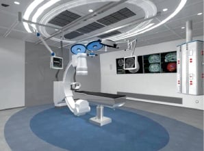
Figure 1. Conceptual animation of the proposed layout for IMRISCardio™ Angio-MR suite in the DR-AR configuration. The fluoro-guided procedure is carried out as usual, and the MR Scanner is only brought into the room when needed. The floor-mounted is swivelled towards a safety area located outside the 50G line of the MR scanner magnetic field.
History
IMRISNeuro™ intraoperative imaging system was conceived as a collaboration between the Department of Neurosurgery at the Foothills Hospital in Calgary, Canada, and researchers at the National Research Council’s Institute for Biodiagnostic, in Winnipeg, Canada. The objective of this project was to develop intra-operative imaging during neurosurgical procedures. IMRIS is today a global leader in intraoperative MR technology for neurosurgical applications, with more than 10 years of clinical experience. Its system is currently marketed worldwide and has been used in a significant number of neurosurgical procedures to date, including CNS neoplasias, epilepsy, cervical spine disorders, arteriovenous malformations, cavernomas and aneurysms. In many cases, the procedure was significantly altered by intra-operatively acquired MR images1 (Figure 2).
Pre-operative image
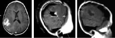
Intra-operative image
Residual tumor
Final check
Figure 2. MR T1-weighted images with Gadolinium contrast agent of a nine year old girl with a giant-cell astrocytoma. Image on the left is a surgical planning scan; image in the centre intra-operatively, in which residual tumour was identified. Image on the right is a post-resection confirmation prior to wound closure. Images courtesy of Dr Garnette Sutherland Calgary Foothills Hospital, Calgary, Alberta, Canada.
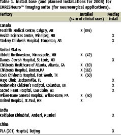
Currently, IMRIS has seven systems installed in North America and eight more installations ongoing, including one in India (Mumbai) and China (Beijing) (Table 1).
Leveraging this experience, IMRIS aims to develop its expertise in interventional imaging in the field of Interventional Cardiology.
With dedicated efforts and a commitment to innovation, IMRIS is developing leading technological solutions for both cardiac and endovascular applications.
Technical specifications
IMRISCardio™ Angio-MR System has the unique ability to move between rooms on demand in a matter of seconds, which provides hospitals with a flexible and cost effective use of these valuable assets. The system allows the surgical team to position the patient exactly as required for the procedure without any compromises. Once positioned, the patient does not need to be moved for intra-procedural MR imaging.
System layout and configurations
Several configurations are possible: DR-AR, AR-DR-OR, AR-DR-AR, (where DR=Diagnostic Room, AR=Angiography Room and OR= Operating Room [Figure 3]).
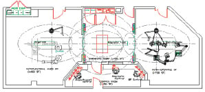
Figure 3. Architectural set drawing for an AR-DR-OR configuration. In this hybrid configuration, the MR scanner serves a cath lab and a surgical room, which are separated by a diagnostic room, where standard diagnostic imaging can also be performed.
In the AR-DR-OR configuration, the MR scanner serves three adjacent rooms designed for different applications: interventional cardiac and endovascular applications, intra-operative (surgical) applications and diagnostic imaging. This provides the hospital with three key major benefits: patients are not required to be moved or repositioned to undergo MR imaging during the procedure; the workflow is minimally impacted, since the scanner is there only when needed; and the ability to serve as a diagnostic tool provides the financial utility of bringing additional revenue to the institution.
System components
IMRISCardio™ Angio-MR System consists of the following components:
– IMRIS MR 15 Imaging System
– Complete Angiography System
– Information Management System (IMS)
– MR-compatible angiography table and coils
IMRISCardio™ MR15 Imaging System
IMRISCardio™ MR15 Imaging System leverages all functionality of the Siemens Espree 1.5 Tesla scanner and combines this with its proprietary magnet mover technology that allows for interventional imaging. The high field 1.5T MR provides the interventionist with sharp, timely baseline and intra-procedural images for pre- and post-intervention evaluation.
Angiography system
IMRIS has extended its partnership with Siemens to include the Artis Zee systems in its integrated IMRISCardio™ imaging suite. The Zee is an all-digital, ceiling or floor mounted angiography system with flat detector technology, designed for diagnostic and interventional procedures. It includes a flexible C-arm positioning around the patient, excellent image quality through CLEAR technology and radiation reduction strategies with CARE applications2. The ceiling mounted gantry has a motorised pivot of 270° providing one of the highest amounts of positioning flexibility for optimised patient access.
Information Management Suite (IMS)
The Interventional Information Management Suite is an integrated operating room specifically designed to provide advanced digital, audio and video capabilities. Image data/video is transferred over a digital architecture from any equipment connected to the system and routes it to any display included in the display set. Since data are transmitted over fibre optic cables, images maintain their native resolution. Its transmission does not interfere with the MRI scanner. Each system is customised according to the type of configuration choosen.
iCMR Table
IMRISCardio Angio-MR table is an MR-compatible angiography table with a metal-free, long cantilever tabletop for MR compatibility, radiolucency, C-arm accessibility and high stability. It offers 4-way motorised tabletop movement, XY translation, height adjustment, Trendelenburg, reverse Trendelenburg, and R/L lateral roll.
iCMR Coils
The system consists of 76 integrated coil elements, which can be combined for 32 channel reception making the system ideals for parallel imaging. The system has a total of 205 cm FOV along the magnet’s long axis, allowing for imaging the entire body without repositioning the patient. The coils provided include:
– Body Matrix Coil – anterior portion of the cardiac coil
– Spine Matrix Coil – for body imaging in the diagnostic room
– IMRIS Cardiac Coil – removable posterior coil for interventional applications
Applications in interventional cardiology
The benefits of intraoperative imaging demonstrated in neurosurgical interventions3,4 can also have a significant impact on patient outcomes when extended to the Interventional Cardiology.
Magnetic resonance is the most suitable imaging modality to complement x-ray fluoroscopy in the interventional cardiovascular setting. MRI allows for excellent tissue delineation and provides information on cardiac anatomy, myocardial function, perfusion, contractility, and physiology without the detriments of ionising radiation. It is suitable to assess a broad variety of cardiac pathologies5, and can provide invaluable information when used during coronary interventions, ablation procedures and other interventions for the treatment of congenital malformations, peripheral vascular and neurovascular disease.
Advanced applications and future opportunities
Near real time and real-time MR guidance is currently being practised and investigated for several applications in cardiovascular medicine. Mainstream areas of Interventional Cardiology in which MR can be of significant value in the future include intravascular imaging for the treatment of coronary artery disease, MR-guided catheter placement and in-vivo lesion visualisation for electrophysiology applications.
Applications in interventional electrophysiology
MRI offers several specific practical advantages over other imaging modalities for guiding and monitoring therapeutic EP interventions:
– Real-time catheter placement with detailed endocardial anatomic information;
– Rapid, high-resolution, 3D visualisation of cardiac chambers;
– High-resolution functional atrial imaging to evaluate atrial function and flow dynamics during therapy;
– The potential for real-time spatial and temporal lesion monitoring during therapy;
– Potential for reduction and eventual elimination of patient and physician radiation exposure.
Applications of IVMRI in coronary artery disease (CAD)
INTRAVASCULAR IMAGING (IVMRI)
Imaging coils can be mounted on guide wires commonly used for angioplasty procedures. MR-based fibrous cap characterisation and measurement in large sections of the coronary arteries (3-6 cm) would be then the best method to determine the need for placement of a vulnerable-plaque specific stent and its long-term follow-up as well as a research tool for the monitoring of other therapeutic modalities currently under development.
INTRAVASCULAR ASSESSMENT OF THE “VULNERABLE PLAQUE”
Catheter angiography shows the plaque as a reverse image but gives no information on plaque structure or plaque components. The composition of atherosclerotic plaques governs their vulnerability to disruption and hence their propensity to cause cardiovascular events6. Current imaging techniques used in clinical practice, such as intravascular ultrasound (IVUS), can delineate plaque size but not content nor structure. MRI can differentiate tissue content within atheroma on the basis of imaging algorithms to optimise contrast.
IN-STENT LATE THROMBOSIS: STENT MALAPPOSITION
Assessment of post-deployment stent-wall apposition has become a significant parameter7 in order to assess the performance of different types of stents. A high-resolution imaging modality such as IVMRI integrated onto the same guide wire that is used to place the balloon and the stent has the potential to become the standard imaging modality for the evaluation of such outcomes.
Development roadmap
Development of the iCMR program is divided in sequential and progressive stages:
Stage 1. Integration of Angio, MR, and IMS
Stage 2. Integration of imaging software
Stage 3. Enhanced interventional MR applications
STAGE 1. ESTABLISHING AN INTEGRATED ANGIO-MR SYSTEM
The main goal in Stage 1 is to achieve optimal performance of all components and sub-systems within the integrated system, both at hardware and software level.
STAGE 2. INTEGRATION IMAGING SOFTWARE
Stage 2 will focus on evaluating dynamic, real-time imaging processing, volume rendering, image-fusion and visualisation algorithms, as well as image registration capabilities, which are required to take full advantage of the synergies between these two imaging modalities.
STAGE 3. ENHANCED INTERVENTIONAL MR APPLICATIONS
Stage 3 is designed to serve as the enabling platform that will allow for the testing of catheters and devices that are MR-safe, compatible and visible. These include passive and active-guidance devices, which are of pivotal importance to visualise the precise location and orientation of the catheter tip, in real time, and with reference to the tissue background.
Conclusion
The possibilities offered by the combination of the two modalities provide exciting new opportunities and advances in cardiac and neurovascular intervention in general, and in the endovascular/ interventional setting in particular. The combined MRI-Angio suite will provide an environment for the development of new therapeutic strategies and devices, by exploring the total integration of the two modalities, each independently representing the state-of-the-art in technological performance.
Supplementary data
To read the full content of this article, please download the PDF.
Moving image 1.