Flashlight

DOI: 10.4244/EIJ-D-25-00121

Calcium mapping by 3DStent technology

Nicolas Amabile, MD, PhD; Hakim Benamer, MD

A 78-year-old man with stable angina, a positive stress test, and a tight left anterior descending artery (LAD) with a mildly calcified lesion on computed tomography (CT) scan (Moving image 1) was referred for coronary angiography. The angiography confirmed the stenosis, and percutaneous coronary intervention (PCI) of this lesion was proposed.

We analysed the calcium burden with a multimodal approach to plan the intervention and the plaque preparation. After initial angiography (Moving image 2), we performed X-ray-based calcium (Ca2+) mapping through adaptation of the 3DStent technology (3DS; GE HealthCare) to the native vessel1. A deflated 2.0x15 mm PCI balloon was first placed within the target lesion, and a 200° rotational angiography was acquired (Supplementary Figure 1). The data were processed through dedicated software which produced 0.1 mm voxel-based images that were visualised with different standard rendering methods, including a three-dimensional image that was analysable in different projections (Supplementary Figure 1, Moving image 3) and cross-section views (Figure 1A). These images provided two- and three-dimensional representations of the calcifications...

Sign in to read
the full article

Forgot your password?
No account yet?
Sign up for free!

Create my pcr account

Join us for free and access thousands of articles from EuroIntervention, as well as presentations, videos, cases from PCRonline.com
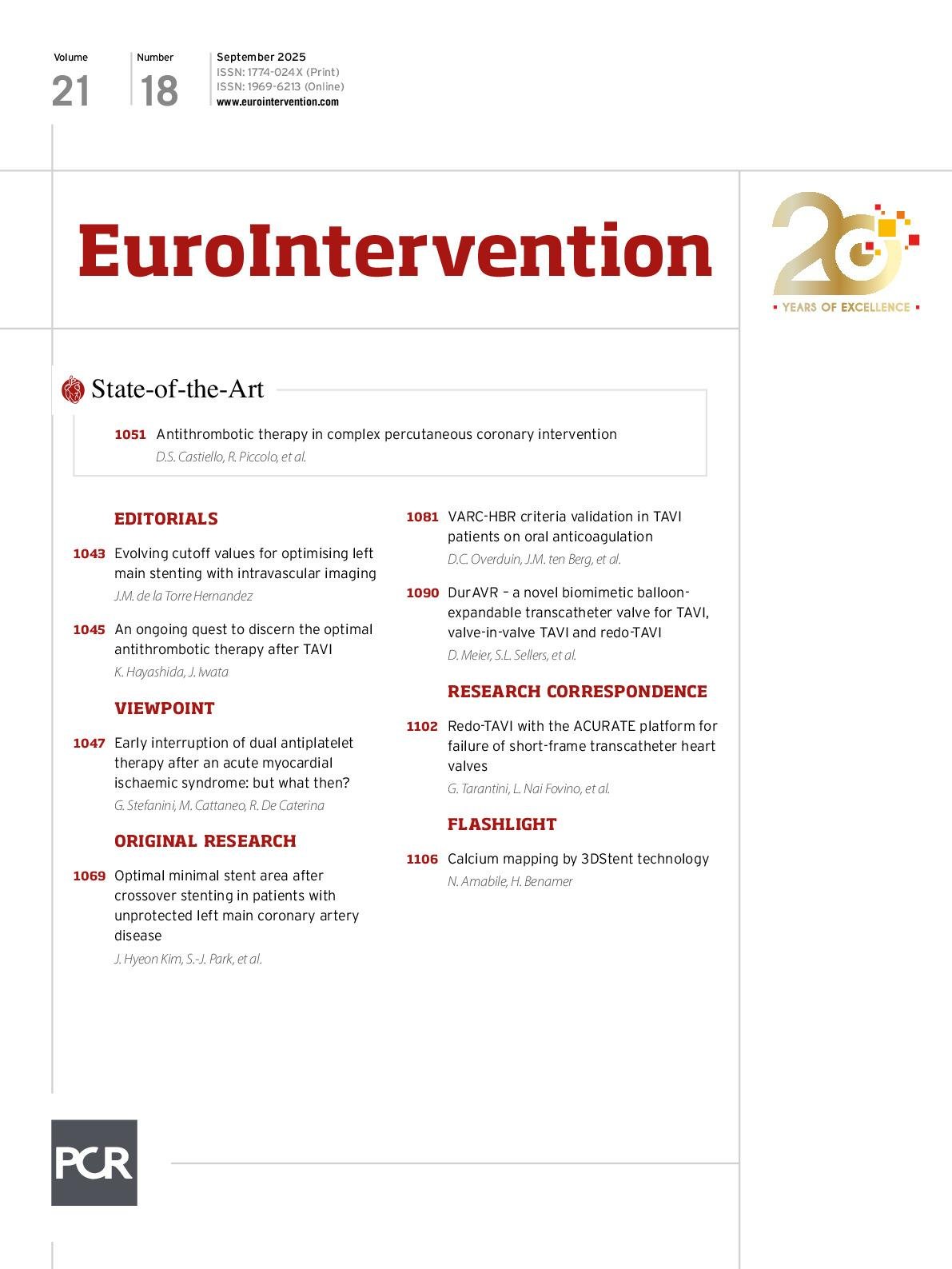
Volume 21 Number 18
Sep 15, 2025
Volume 21 Number 18
View full issue


Key metrics

Suggested by Cory

CLINICAL RESEARCH

10.4244/EIJ-D-17-00962 Apr 6, 2018
A new optical coherence tomography-based calcium scoring system to predict stent underexpansion
Fujino A et al
free

State-of-the-Art

10.4244/EIJ-D-24-00195 Apr 7, 2025
Percutaneous coronary intervention for calcified and resistant lesions
Pesarini G et al

State-of-the-Art

10.4244/EIJ-D-24-00387 Jul 21, 2025
Advances in coronary imaging of atherosclerotic plaques
Garcia-Garcia H et al
free

CLINICAL RESEARCH

10.4244/EIJ-D-16-00139 Jun 20, 2017
Three-dimensional volumetric assessment of coronary artery calcification in patients with stable coronary artery disease by OCT
Krishnamoorthy P et al
free

Debate

10.4244/EIJ-E-23-00002 Jun 5, 2023
All calcified nodules are made equal and require the same approach: pros and cons
Guagliumi G et al
free

Expert review

10.4244/EIJ-D-18-01056 Oct 18, 2019
Intravascular lithotripsy for calcific coronary and peripheral artery stenoses
Dini C et al
free
Trending articles
76.3

State-of-the-Art

10.4244/EIJ-D-23-00840 Sep 2, 2024
Aortic regurgitation: from mechanisms to management
Baumbach A et al
free
56.1

Original Research

10.4244/EIJ-D-25-00331 May 21, 2025
One-month dual antiplatelet therapy followed by prasugrel monotherapy at a reduced dose: the 4D-ACS randomised trial
Jang Y et al
open access
56.1

Original Research

10.4244/EIJ-D-25-00331 Jul 21, 2025
One-month dual antiplatelet therapy followed by prasugrel monotherapy at a reduced dose: the 4D-ACS randomised trial
Jang Y et al
open access
31.1

Expert Review

10.4244/EIJ-D-24-00535 May 5, 2025
Catheter-based techniques for pulmonary embolism treatment
Costa F et al
28.25

Clinical research

10.4244/EIJ-D-19-01006 Aug 7, 2020
Coronary collaterals and myocardial viability in patients with chronic total occlusions
Schumacher S et al
free
X

PCR
Impact factor: 9.5
2024 Journal Citation Reports®
Science Edition (Clarivate Analytics, 2025)
Online ISSN 1969-6213 - Print ISSN 1774-024X
© 2005-2025 Europa Group - All rights reserved