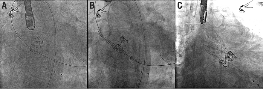
Percutaneous implantation of an aortic valve is an alternative for the treatment of bioprosthetic surgical valve deterioration. Less is known about the treatment options for failed catheter implanted valves. We report herein a case of an early degeneration of the Edwards SAPIEN XT balloon-expandable valve (Edwards Lifesciences, Irvine, CA, USA) which we treated using the successful implantation of a self-expandable CoreValve® (Medtronic, Minneapolis, MN, USA) in a patient sustaining haemodialysis. The patient was referred to our service due to signs and symptoms of severe aortic stenosis 20 months after transcatheter aortic valve implantation (TAVI) using the balloon-expandable Edwards SAPIEN XT 23 mm device. Echo findings showed thickened and poorly mobile aortic valve leaflets, and pressure gradients were 90/60 mmHg. Following consideration of the treatment options, we performed a second valve-in-valve TAVI procedure using a self-expandable CoreValve Evolut 23 mm device (Medtronic). The procedure was carried out smoothly and without complications (Figure 1, Online Figure 1, Moving image 1-Moving image 6). The valve residual gradient was 28/18 mmHg and without residual paravalvular leak. This case indicates a possible relationship between the earlier catheter-based valve degeneration and the renal insufficiency/dialysis treatment characterising our patient. Importantly, it may also point towards an expansion of the future treatment options following late failure of catheter-based valve devices.

Figure 1. CoreValve Evolut 23 mm within Edwards SAPIEN XT 23 mm. A) A stiff guidewire has been crossed to the left ventricle through the Edwards SAPIEN XT valve. B) The self-expandable CoreValve within the Edwards SAPIEN XT in the pre-deployment phase. C) Final position of the self-expandable device within the balloon-expandable valve.
Conflict of interest statement
The authors have no conflicts of interest to declare.
Online data supplement
Moving image 1. Pre valve-in-valve echo.
Moving image 2. Pre valve-in-valve angio 1.
Moving image 3. Pre valve-in-valve angio 2.
Moving image 4. Deployment landing angio.
Moving image 5. Deployment completion angio.
Moving image 6. Post valve-in-valve angio.

Online Figure 1. Haemodynamic tracing pre- and post-deployment.
Supplementary data
To read the full content of this article, please download the PDF.
Moving image 1. Pre valve-in-valve echo.
Moving image 2. Pre valve-in-valve angio 1.
Moving image 3. Pre valve-in-valve angio 2.
Moving image 4. Deployment landing angio.
Moving image 5. Deployment completion angio.
Moving image 6. Post valve-in-valve angio.

