I’ve always envied centres with a well-established, well-attended Journal Club. I became aware of this format several years ago during my American fellowship period, and since then I have always wanted to import it into my centre. The formula is simple: an hour, a recent (perhaps controversial) paper, a faculty in the mood for stimulating criticism, and curious fellows.
The first problem was establishing the time for the meeting. In the middle of the morning? Impossible, everyone is too busy. At the end of the morning? Maybe too tired? Early in the morning? Too lazy, or maybe not. That’s where the beauty lies, in making a small sacrifice to take care of yourself, and in this case of your own mind and critical thinking. It’s no different than when you go to the gym on a rainy day, even though you want to use the rain as an excuse to stay home.
So, the Journal Club is held at 7:15 in the morning. Which discourages casual attendees but is also motivating for the well intentioned. We started over a year ago (I’m writing to you during the second season), and there were only four or five of us. The format is a reinvention of the original: a junior fellow prepares a 10-minute presentation of the selected paper; the senior fellow prepares a 10-minute critique of the selected paper; the remaining participants (faculty and fellows) comment on what they saw during the next 20 minutes (more or less, because the only goal is to finish before 8:00, when the hospital comes alive).
The beauty of this format is that it always starts, inevitably, at 7:15, even if the world is falling apart. It is so predictable (it never misses an appointment) that it can even outlive its main actors. It’s now a habit, every other Tuesday, like coffee. The fellows rotate, and now they even spontaneously offer to present something themselves. The hospital audience has grown by word of mouth (Instagram was instrumental in attracting the youngest), and now, sometimes we can’t all fit into our classroom (depending, of course on the topic). The discussion is formidable, and the main cardiology journals offer us exciting new topics every two weeks. Of the small academic accomplishments that I have achieved in my life, this one gives me special satisfaction. It doesn’t take much and can be done anywhere. The ingredients are enthusiasm, people who share in this spirit, and an article.
Sometimes an article is from EuroIntervention, and it might even be included in the ones I’m presenting now in this issue!
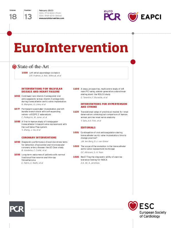
This month, we begin with a State-of-the-Art by David R. Holmes Jr, Mohamad Alkhouli and colleagues on left atrial appendage occlusion. While oral anticoagulation is a cornerstone of atrial fibrillation-related stroke prevention, the number of non-valvular atrial fibrillation patients has risen over the last few decades. For non-valvular atrial fibrillation patients, the aetiology of cardioembolic stroke is, in 90% of cases, thrombus in “the most lethal human attachment”, the left atrial appendage. This review focuses on the pathophysiology, patient selection, procedural performance, treatment outcomes, adjunctive therapy, complications, and longer-term outcomes of left atrial appendage occlusion for selected patients with non-valvular atrial fibrillation at risk for stroke.
In interventions for valvular disease and heart failure, Norman Mangner, Axel Linke and colleagues report on whether continued non-vitamin K antagonist oral anticoagulants during TAVI are as safe as continued vitamin K antagonists. At 30 days, they found comparable outcomes with the primary composite outcome measures of major/life-threatening bleeding, stroke and all-cause mortality. This article is accompanied by an editorial by Jurriën M. ten Berg and Dirk-Jan van Ginkel.
Next in interventions for valvular disease and heart failure, Costanza Pellegrini, Michael Joner and colleagues look at the development of new conduction disturbances (i.e., left bundle branch block) and the potential need for new permanent pacemaker implantations post-TAVI in a subanalysis of the SCOPE II trial. Here, they assess the incidence and impact of new left bundle branch block and permanent pacemaker implantations (PPI), using the ACURATE neo and the CoreValve Evolut R, both of which are self-expanding transcatheter heart valve devices. They found lower rates of both PPI and left bundle branch block with the ACURATE neo as well as an associated decreased risk of PPI with the ACURATE neo device. This article is accompanied by an editorial by Guilherme F. Attizzani and Sung-Han Yoon.
Finally, in interventions for valvular disease and heart failure, a research correspondence by Yuan Zhang, Junbo Ge and colleagues reports on a first-in-human study using the LuX-Valve Plus, a new-generation of the LuX-Valve, specifically adapted for transjugular transcatheter tricuspid valve replacement. Patients with symptomatic severe tricuspid regurgitation at high surgical risk were included, and the device was successfully implanted in all patients. At 30 days, all patients had none/trivial tricuspid regurgitation.
Coronary microvascular dysfunction may affect the results of non-invasive tests. In the section on coronary interventions, Bert Vandeloo, Carlos Collet and colleagues report the results of the UZ Clear study which included patients with an intermediate pre-test probability of coronary artery disease and positive exercise stress tests referred for invasive angiography. The objective was to determine the false discovery rate of cardiac exercise stress tests with both fractional flow reserve (FFR) and the index of microvascular resistance as references, with the combination of these two references as clinical standards reducing the false discovery rate by 25% compared to quantitative coronary analysis. An invasive functional assessment accounting for the epicardial and microvascular compartments led to an improvement in the diagnostic performance of exercise tests. This article is accompanied by an editorial by Ziad A. Ali and Allen Jeremias.
Also in coronary interventions, in an extended follow-up of the COMBINE OCT-FFR study, Enrico Fabris, Elvin Kedhi and colleagues look at the long-term outcomes of patients with diabetes mellitus, normal FFR and thin cap fibroatheroma. Thin cap fibroatheroma-positive lesions were associated with a higher risk of adverse events during long-term follow-up, and the recurrence of target lesion-related major cardiovascular events was higher in thin cap fibroatheroma-positive patients beyond 18 months and up to five years. Optical coherence tomography-guided detection of high-risk lesions can help determine which patients may need a more aggressive treatment strategy.
Next, Giuseppe Tarantini, Francesco Burzotta and colleagues investigate the safety and efficacy of left main percutaneous coronary interventions using the Resolute Onyx drug-eluting stent from the ROLEX Registry. This latest-generation zotarolimus-eluting coronary stent includes dedicated extra-large vessel platforms. Target lesion failure at one year was low, and the primary endpoint incidence was significantly lower in patients undergoing intravascular ultrasound/optical coherence tomography-guided versus angio-guided left main stenting, though further randomised trials are called for when using drug-eluting stent platforms without dedicated large-vessel designs.
In the section on interventions for hypertension, Yu Sato, Aloke V. Finn and colleagues offer this translational research examining the histology in normotensive porcine treated with radiofrequency renal denervation compared to untreated human cadavers. The distribution of nerves and the relative distribution of peri-arterial structures was similar between humans and porcine, validating the translational value of the normotensive porcine model for renal denervation.
And now, let’s let the articles speak for themselves.

