Abstract
Background: After extensive in vitro and animal testing, we performed the first retrograde transcatheter implantation of a new aortic valve prosthesis, in a 62-year-old man with inoperable calcific aortic stenosis.
Methods and results: A 16 F sheath was advanced into the abdominal aorta via the right femoral artery. The aortic valve was crossed with a straight wire, and a pigtail catheter was advanced into the left ventricle to obtain pressure-gradient and anatomic measurements. Predilatation was done with an 18-mm valvuloplasty balloon. A balloon-expandable frame was used to deliver the valve. After device implantation, transvalvular gradient was <5 mmHg. Cardiac output increased from 1 to 5 L/min, and urine production increased to 200 mL/h. The patient was extubated on the second post-implant day. Twelve hours later, he had to be re-intubated because of respiratory distress and high pulmonary pressures. His condition deteriorated, and he died on day 5, of biventricular failure and refractory hypotension. Despite the severe hypotension, valve function was satisfactory on echo-Doppler evaluation.
Conclusions: Retrograde transcatheter implantation of a prosthetic aortic valve yielded excellent haemodynamic results and paved the way for further use of this technique in selected high-risk patients.
Efforts to develop a percutaneously implantable heart valve date back to 1965, when Davis1 described an umbrella-like device for relieving experimentally induced aortic insufficiency in dogs. Since that time, other researchers2-11 have published their experiences with percutaneous valve implantation in animal models. In 2002, Bonhoeffer and his group12,13 performed the initial human percutaneous valve implant in the pulmonary position. That same year, Cribier and co-workers14 described the first clinical antegrade transcatheter placement of an aortic valve.
We report the first retrograde transcatheter implantation of an aortic valve, in a 62-year-old man with inoperable calcific aortic stenosis, intractable congestive heart failure, severe pulmonary hypertension, renal failure, and passive liver congestion.
Materials and methods
In vitro evaluation
Over the past 9 years, we have developed a biologic valve15 (the Paniagua Heart Valve [PHV]; Endoluminal Technology Research, Miami, Florida) that has a collapsed profile of 2 mm that can be inserted with an 11F or a 16F introducer, depending on the mounting frame and final valve diameter. These characteristics are highly suitable for retrograde transcatheter delivery.
The PHV was evaluated in a left-heart and systemic circulation simulator (Vivitro Systems, Victoria, Canada). This system includes a processor-controlled stepper motor that drives a piston cylinder, forcing contraction and relaxation of the left ventricular sac. The aortic valve was mounted in its relative anatomic position. To achieve the same density and viscosity as blood, the system was filled with a mixture of 70% water and 30% glycerol. Sodium chloride (0.9%) was added to allow flow to be measured with an electromagnetic probe. The PHV was tested under physiologic conditions (cardiac output, 5 L/min; heart rate, 70/min; blood pressure, 180/80 mmHg) and pathologic states of hypotension (70/40 mmHg) and hypertension (320/250 mmHg). Echo-Doppler studies were performed with a 7.5-MHz transducer in the in vitro model. The PHV had no transvalvular gradient and no backflow (regurgitation) under physiologic conditions. Its opening gradient was 5 mmHg. Valve opening and closing characteristics were excellent. During extreme hypertension (320/250 mmHg), no prolapse was seen. Two-dimensional echocardiographic images showed complete apposition of the leaflets and the typical Mercedes-Benz sign. Pulsed Doppler studies of the acute in vitro model showed physiologic forward flow velocities and no evidence of regurgitation.
We tested the PHV in a long-term in vitro model for more than 2 years (236 million opening and closing cycles) and observed no device dysfunction or deterioration in materials.
Animal studies
In 2000, we began a series of short- and long-term animal experiments, some of which are ongoing. The initial studies were performed in Costa Rica and Miami Beach, Florida, using sheep and calf models. All the animals were treated humanely, according to the standard procedures for animal research used at the Biomedical Research Institute, Mount Sinai Medical Center, Miami Beach, Florida. Eleven sheep and 6 calves, weighing 30 to 60 kg each, underwent transcatheter implantation of the PHV. Eight chronic and 9 acute animal studies were performed.
Vascular access was achieved percutaneously or by surgically exposing a peripheral artery (femoral or carotid). The arterial entry site was enlarged with dilators. Baseline and post-implant angiograms were then obtained. The PHV was implanted in the descending aorta of 11 animals and in the ascending aorta of the other 6 animals. The prosthesis was introduced with a customized delivery system and was deployed in the selected anatomic position under fluoroscopic guidance. A balloon-expandable stent was used in 1 case, and a self-expanded stent was used in the other 16 cases; all stents were oversized by 10% in order to exert enough radial force to hold the valve in place without migration. Prophylactic antibiotics were given. Once the valve was delivered, its position and function was verified with transthoracic Doppler ultrasonography. Complications occurred in 2 cases: in 1, the stent failed to open, causing fatal aortic thrombosis. In the other case, the animal died suddenly of postoperative vascular complications at the access site. No migration of the stented valves occurred. One calf was sacrificed 3 weeks after PHV implantation. Pathologic examination of the implant site showed ecchymoses and superficial hemorrhage without evidence of perforation or dissection. The harvested prosthesis functioned competently in the left-heart simulator and showed no histologic evidence of rejection. One calf was sacrificed 13 months after implantation of a self-expanding stent in the descending thoracic aorta. A CT scan of the valve showed no calcification. Six sheep were sacrificed 6 months post-implant. The explanted PHVs showed endothelialization without an inflammatory reaction. At sacrifice, all of the animals were in excellent condition.
Clinical case
A 62-year-old man had a history of infrapopliteal peripheral vascular disease and severe aortic stenosis, which evolved into severe pulmonary hypertension, biventricular failure, passive liver congestion, and chronic renal failure (baseline creatinine level, 2.7 mg/dl). He was admitted to Centro Medico de Caracas Hospital with anasarca, severe pulmonary œdema, left pleural effusion, pulmonary hypertension, right-sided heart failure, hepatomegaly, ascites, and significant scrotal and lower-limb œdema. Despite aggressive diuretic and inotropic support his condition failed to improve, and oliguric renal failure developed.
Transthoracic echocardiography showed global hypokinesis with an ejection fraction of 15%, an apical thrombus, a peak aortic gradient of 50 mmHg, and an aortic valve area of 0.6 cm2. The native valve showed eccentric calcification with decreased opening excursion and fusion of the three leaflets. The pulmonary pressure was 60 mmHg, based on the tricuspid regurgitant jet and a presumed right atrial pressure of 10 mmHg. The coronary arteries had no significant disease, and the native aortic valve area was 0.53 cm2 according to the Gorlin formula. Because of the patient’s low ejection fraction, comorbidities, and generally hopeless situation, his case was declined by 3 surgical groups. He was offered a PHV by his treating cardiologist, and the case was discussed at length with the family. They understood the procedure’s risks and benefits, as well as the alternative options, and they wished to proceed. The hospital’s institutional review board approved the procedure for compassionate use as a possible life-saving strategy.
In the cardiac catheterization laboratory, the patient underwent mild sedation and local anesthesia of both groins. A 6F catheter was advanced from the left femoral artery to the ascending aorta to allow initial coronary angiography and continuous blood-pressure monitoring. Two sheaths were placed in the left femoral vein; 1 sheath for right-sided heart catheterization and the other for placing a temporary pacemaker in the right ventricle. The right femoral artery was surgically exposed to allow optimal control of the access site and to prevent local complications related to the severe peripheral vascular disease. A 16F Cook introducer (Cook, Inc., Bloomington, Indiana, USA) was advanced into the abdominal aorta. A straight wire was then used to cross the aortic valve, and a calibrated pigtail catheter was advanced into the left ventricle. The transvalvular peak gradient was 36 mmHg. An Amplatz super-stiff wire (Medi-tech/Boston Scientific, Natick, MA) was positioned in the left ventricle through the pigtail catheter. Intravascular ultrasonography was done with a 20-MHz, 6F catheter and a Galaxy ultrasound imaging system (Boston Scientific). By means of planimetry, we estimated that the aortic valve area was 0.6 cm2. An 18-mm-diameter valvuloplasty balloon (Scimed, Boston Scientific), measuring 6 cm in length, was placed in the aortic valve and fully inflated. To deliver the device, we used a CP 8Z28 Stent (NuMED, Hopkinton, NY) and a 20-mm balloon-in-balloon catheter. The 20-mm chemically sterilized PHV was oversized by 10%, to fit the diameter of the left ventricular outflow tract. The prosthesis was manually crimped to a diameter of 4 mm and was then advanced over the wire to the aortic valve plane.
During placement of the valve, the patient had a cardiac arrest, which was probably related to aortic outflow obstruction. The PHV was withdrawn into the aortic arch, and cardiopulmonary resuscitation was initiated. Sinus rhythm was restored in less than a minute, and the patient had no neurologic sequelae. He was intubated and kept comfortable with mild general anesthesia. By using fluoroscopic landmarks and overlapping images of the right coronary ostium, both in the same 40-degree left anterior oblique orientation with 20-degree cranial angulation, we were able to deploy the PHV successfully in the subcoronary position (Figure 1).
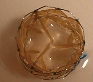
Figure 1. The Paniagua heart valve
We noticed a waist in the PHV in the aortic valve plane but refrained from further attempting to enhance the valve diameter for fear of causing a vascular rupture.
Results
The implantation and fluoroscopy procedures took 130 and 22 minutes, respectively. After the valve was deployed, the patient’s haemodynamic values improved significantly. The aortic pressure increased from 80/50 to 110/60 mmHg, and the transvalvular gradient decreased from 36 to <5 mmHg. Immediately after valve expansion, a complete atrioventricular block occurred, and pacing was required for 45 seconds, after which the heart spontaneously returned to sinus rhythm. The cardiac output increased from 1.0 to 2.8 (later 4.8) L/min, and urine production increased to 200 mL/h.
Imaging studies
Gadolinium-enhanced aortography showed no regurgitation through the PHV. Only a small paravalvular leak was present, and both coronary ostia were patent. A right anterior oblique cranial view showed that the stent had completely expanded in a circular fashion. Intravascular ultrasonography with a 20-MHz probe confirmed excellent expansion of the stent, as well as good mobility and apposition of the PHV leaflets. The PHV was located eccentrically because of severe calcification and post-stenotic dilatation of the coronary sinus and aortic valve leaflet. The diameter of the valve perfectly matched that of the outflow tract and the area of aortic calcification. We used the Gorlin formula16 to calculate the PHV area, although that formula has not been validated for this particular setting. According to the Gorlin formula, the aortic valve area was 1.27 cm2 when the cardiac output was 2.8 L/min, and the valve area was 2.2 cm2 when the cardiac output was 4.8 L/min.
Transthoracic echocardiography, performed within 30 minutes after PHV implantation, revealed a completely excluded native aortic valve and a circular stent geometry with a diameter of 20 mm. Prosthetic valve function was optimal, with a mean gradient of 5 mmHg, a valve area of 1.6 cm2 as measured by planimetry in a cross-sectional view, and mild paravalvular regurgitation between the PHV and the heavily calcified aortic valve leaflets.
Twenty-four hours after device implantation, transoesophageal echocardiography showed satisfactory PHV hemodynamics. Serial echo-Doppler studies confirmed good valve function even when the patient was hypotensive. Nevertheless, the left ventricular ejection fraction remained poor, ranging from 10% to 20%.
Clinical evolution
After valve implantation, permanent anticoagulation therapy was initiated with intravenous heparin and antiplatelet agents (aspirin and clopidogrel). During the first 48 hours post-implant, the patient’s pulmonary and peripheral congestion was dramatically reduced. His urine output increased to 200 mL/h, and he was extubated on the second day. On the third day, however, he developed respiratory distress, and his pulmonary pressure increased to systemic levels (90 mmHg). A pulmonary embolism was suspected, aggressive anticoagulation was continued, and reintubation was required. Within the ensuing 48 hours, biventricular failure was followed by refractory hypotension, and the patient died on post-implant day 5. Despite the severe hypotension, valve function remained satisfactory, as evaluated with echo-Doppler methods. Unfortunately, permission for an autopsy could not be obtained.
Discussion
Ours is the second report of percutaneous valve implantation in the aortic position. However, it is the first case to involve retrograde implantation of such a valve, thereby confirming the feasibility of this approach, which follows standard interventional techniques. In 2000 Bonhoeffer and colleagues,12 performed the first percutaneous replacement of a failed pulmonary valve in a right-ventricle to pulmonary-artery prosthetic conduit.
In the first clinical transcatheter aortic valve procedure, performed in 2002, Cribier and coauthors14 used the antegrade approach because their device required a 24F sheath (outer diameter, 26F), which would have been hard to advance through the arterial system. However, the antegrade approach has several disadvantages, including the need to perform a transseptal procedure with the risk of perforation and cardiac tamponade; the need to create an atrial septal defect; the risk of advancing the wire through the mitral subvalvular apparatus; and the risk of rupturing the mitral valve during balloon inflation, especially if the wire is between the mitral chordae tendineae.
By percutaneously implanting the PHV, we obtained a successful short-term therapeutic result in an otherwise hopeless situation. The mechanism of cardiac arrest that occurred during initial placement of the device in the aortic valve plane may have been related to outflow obstruction with a decreased blood supply to the coronary arteries in a patient with no cardiac reserve. Before valve implantation, the patient’s cardiac output was incompatible with life. After device implantation, the patient recovered impressively. When he was in the intensive care unit, receiving inotropic and vasodilator support, his cardiac output increased dramatically, as did the valve area calculated with the Gorlin equation. The PHV functioned satisfactorily at all times, as confirmed by both transthoracic and transoesophageal echocardiography. Even when the patient was hypotensive, valve opening and closing was adequate.
The waist seen in the PHV after deployment was due to overexpansion of the borders of the stent (both upper and lower). Because there was no gradient across the valve and because intravascular ultrasonography showed good stent apposition to the aorta, no further dilatation was done.
The cause of the patient’s acute collapse after a period of significant improvement is unclear. Antegrade migration of the valve with coronary ostial obstruction is ruled out, because this complication results in sudden death if the device occludes the left main coronary artery or significant electrocardiographic changes if it occludes the right coronary ostium or both arteries. Retrograde migration causes severe mitral insufficiency, which was not seen on echo-Doppler studies. During TEE, the valve was seated in the aortic valve plane, both coronary ostia were visible, and no mitral insufficiency was detected. Across the PHV, 2 m/s jets were accompanied by early peaks indicating a lack of significant obstruction.
Another possible explanation is sepsis, but the patient’s temperature and white blood cell count were normal. Daily blood cultures also yielded negative results. When the patient collapsed, his pulmonary pressure increased from 60 to 90 mmHg. This clinical scenario suggests a pulmonary embolism as the most likely diagnosis. The patient’s condition was too unstable for confirmatory testing. Thrombolytic agents were contraindicated because of the recent need for cardiopulmonary resuscitation.
The optimal anticoagulant regimen for use after PHV implantation still needs to be defined. Currently, it consists of heparin followed by oral anticoagulant and/or antiplatelet therapy.
In the treatment of valvular disease, the percutaneous route is a new approach that can be expected to benefit many patients in the future. At present, this procedure is limited to terminally ill patients with severe aortic valve stenosis not amenable to surgical valve replacement. With time, further device modifications should allow transcatheter treatment of valvular disease to become a widely used technique.
Acknowledgments
The authors thank the personnel of the cardiac catheterization laboratory and intensive care unit at Centro Medico de Caracas, Caracas, Venezuela; and Guillermo Villoria, MD, Francisco Mejia, MD, Maria G. Hernandez, MD, José F. Condado, BSc, and Carolyne Ortiz, BSc, for their invaluable support during the procedure. We also thank Virginia Fairchild for her editorial assistance. We especially thank the patient’s family for their trust in this new technology.