Introduction
In the realm of emerging coronary interventional technologies, an innovative Magnetic Navigation System (MNS) has been developed (Niobe, Stereotaxis, St. Louis, MO) for integrated clinical use with a digital, angiographic coronary system (AXIOM Artis dFC; Siemens, Forchheim, Germany). It has been designed to allow the coronary interventionalist to utilize the technique of Magnetic Assisted Intervention (MAI); whereby the operator can navigate the magnetic tip of a specifically designed 0.014 in. guide wire through the coronary vasculature by magnetically deflecting the wire tip in vivo without the need for a pre-shaped bend. An additional novel technology, which is under development, is the remote navigation system (RNS) designed to allow remote, parallel advancement of both the interventional guide wire and device (NaviCath,LTD,Haifa,Israel). It would allow the operator perform to interventional coronary procedures from a location remote from the fluoroscopic table with a joy-stick/ touch-screen computer interface.
With the advent of increased clinical utility of drug-eluting stents (DES), the decreased restenosis rate and increased durability of these devices compared with standard stents has been well reported1,2. This clinical advancement has allowed increased efficacy in the treatment of complex and multivessel coronary disease. However, effective therapy can be limited by the occasional inability of the interventionalist to navigate the standard available guide wires through anatomically challenging regions of coronary vasculature and atherosclerotic disease. Coronary vessels that are excessively tortuous, calcified, and have angulated branches can lead to technical limitation in reaching and crossing distal, eccentric, and long coronary stenoses. It has been shown experimentally, in a three-dimensional phantom model, that utilization of the MAI technique had increased success in navigating successfully through complex turns. Additionally, both procedure time and fluoroscopy time were significantly reduced by MAI compared with standard wire navigation3. Initial reports on utilization of the MAI technique in animal neuro-vascular4 and human electrophysiology studies5 have shown magnetic navigation to be feasible and safe in those relative experimental and clinical settings. In cardiac electrophysiology, magnetically enabled catheters have been evaluated that have the technical ability of both remote advancement and magnetic tip deflection6. There is currently no clinically available device to allow remote magnetic guide wire (or interventional device) advancement.
In the field of interventional cardiology, K. Tsuchida et al.7 and K. Hertting et al.8 have now independently reported their initial clinical experience, efficacy, and safety in patients undergoing MAI in their cardiovascular interventional laboratories. R. Beyar et al.9 reports on the initial experimental feasibility and safety of remote, parallel advancement of the interventional guide wire and device.
The science of Magnetic Assisted Intervention
The magnetic guidance system is comprised of two focus-field permanent magnets, encased within a durable fiberglass housing, and normally kept in the stowed position laterally opposed to the walls of the coronary angiographic laboratory. The angiographic equipment has been specifically adapted to operate within the magnetic environment with flat screen detectors and monitors being utilized throughout the suite. When activated, the magnets rotate into the navigant position on either side of the fluoroscopic table and become computer-integrated with the digital angiography system. This produces a 15 cm uniform, spherical magnetic field within the patient’s chest region (0.08 Tesla). The permanent magnets are mounted on mechanical positioners, which rotate and translate the magnets to generate, under specified computer control, a specified field direction (net magnetic vector) at the tip of the magnetically-enabled guide wire. Using the computer interface system at tableside, through either the mouse-control or touch-screen technology, the interventionalist can direct the resultant magnetic vector to any orientation in three-dimensional space.
Magnetic navigation results from placing the recently developed guide wire (Cronus: Stereotaxis, St Louis, Mo), which has a 3 mm gold-encapsulated neodymium iron boron magnet at its distal tip, within the magnetic field and allowing the magnetically driven deflection of the distal tip to guide the wire through angulated segments while manually advancing the wire. As described by Tsuchida6, the computer user interface allows the operator a variety of options to manipulate the magnetic vector to achieve optimal vector orientation. These initial clinical reports were described utilizing the first two clinically available magnetic guide wires: Cronus Floppy and Moderate-Support wires.
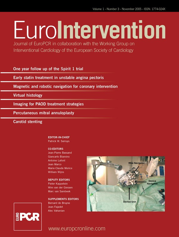
Although not currently associated with distal magnetic navigation, the remote navigation system is composed of a bedside advancement unit that is designed to accommodate both the guide wire and balloon/stent delivery system catheters; and a remote manipulation unit. The computer interface allows both axial and rotational guide wire manipulation. The reported pre-clinical study uses conventional guide wires and interventional devices.
Critique
In regards to the initial clinical application of MAI within the coronary interventional patient population at the Thoraxcenter at Rotterdam, Tsuchida reports the results of the first 59 patients (68 target lesions). This is an observational study of non-consecutive patients who were selected for MAI. The patients appear to have been selected by physician preference and not by specified clinical or lesion-characteristic criteria. The patients were excluded for acute myocardial infarction (AMI), visible thrombus, claustrophobia, or renal insufficiency. No chronic total occlusions (CTO) were included in this group. Procedural success was defined as navigating the magnetic guide wire passed the coronary stenoses without procedural complication.
Successful magnetic guidance was reported in 88% (60/68) of coronary lesions. Four of the unsuccessfully crossed stenoses were characterized as short/eccentric, one as eccentric, and two as poorly visualized secondary to vessel overlap. The only complication noted was one minor dissection, for which MAI was aborted, and a stent was subsequently successfully placed.
Of significant clinical interest, 7/8 coronary stenoses unable to be crossed with the MAI technique could be crossed with conventional wires. Conversely, 4 stenoses that had been previously delineated as being unable to be crossed with conventional guide wire technique were successfully crossed with MAI. One was a distal SVG stenosis with proximal tortousity and 3 were side-branch vessels “jailed” by previously placed stents. It is also noted that in only 19 lesions (28%) was the magnetic wire utilized for PCI.
This study is purely observational and is influenced by investigator selection bias for patients selected for both primary and secondary MAI approach. In this cohort of patients, the technique was found to be unsuccessful in 10% of patients, who subsequently achieved a successful navigational result with the use of a conventional guide wire. Conversely, MAI was successful in four randomly selected cases where conventional wires had previously failed. The magnetic navigation technique was considered safe with only one minor dissection reported.
The initial clinical experience reported by Hertting from Allgemeines Krankenhaus St. Georg in Hamburg, has shown a lower MAI success rate with 63/82 lesions (77%) being successfully crossed. Of the remaining 19 lesions, 13 were successfully navigated with conventional wires and the remaining 6 could not be crossed. Patients were excluded if they had AMI, ICD, or PPM. The clinical populations, however, are not totally comparative as 10 lesions in this group were CTOs, which had been excluded in the Rotterdam group. If one excludes the 2 unsuccessful CTO procedures and the 10 CTO lesions, then the success rate for subtotal coronary lesion navigation was 61/72 (85%), which similar to the 88% success rate reported by Tsuchida. The authors also admit that the observed success rate may have been influenced by the degree of difficulty among chosen cases, with 50% being type B2/C lesions. Additionally, the data also includes 6 lesions that previously were unsuccessfully crossed by conventional wire technique, of which 3 (50%) were successfully crossed with MAI. Thereby showing that in this cohort as well, there were a small group of patients where MAI enabled a previously unsuccessful PCI. Lesion characteristics most associated with failure of magnetic navigation were sited as severe calcification5 or severe tortuosity5.
Of clinical note is the higher complication rate of this study patient group compared with the Rotterdam study. There were five major dissections and 1 death reported. However, of these it appears only 3 dissections, during attempted magnetic wire crossing of a subtotal lesion, were directly related to the MAI technique with the other complications secondary to guide catheter or PCI device trauma. Again, given that patient selection and therapy were guided by physician preference, and not a prospective investigational protocol, one must treat these outcomes as observational only.
In their pre-clinical experiments in Haifa, Beyar describes initial utilization of RNS in an in vitro glass coronary model and in vivo sheep model of normal coronary vasculature. Within their phantom model, it was found that the wire could easily be remotely navigated to each desired location and the interventional device successfully positioned at the desired position. The angulations of the branches within the model are not discussed and therefore the degree of geometrical challenge to remotely navigating through the model cannot be evaluated. The animal model was utilized to investigate the remote navigation technique in normal coronaries with 7 different conventional guide wires and was reported as successful in each trial at reaching the desired location. Bifurcations and regions of tortuousity were successfully crossed but without discussion on the degree of angulations. There is also no reference to the extent that the distal guide wire was pre-shaped (distal tip angulation) and whether the techniques of re-shaping the distal tip or wire exchange was necessitated for successful navigation. Interventional devices were successfully placed at desired locations in 12/14 (85.7%) attempts. Failures were attributed to prototype device malfunctions and not to anatomic coronary challenges.
Safety considerations
In a discussion of the safety and feasibility of the MAI technique, one must emphasize the importance of the adoption of an institutional magnetic safety protocol coincidental to the instillation of a magnetic navigation system. Although the field strength is considerably less than a comparable MRI imaging system (0.08 Tesla vs. 1.5 Tesla), patients should be routinely screened for magnetic exclusions in a similar fashion to that performed before an MRI procedure. Ocular metallic particles, cerebral vascular clips, and permanent pacemakers are important to identify prospectively. Patients who are pacer-dependent or have ICDs should be excluded. Patients with non-dependent permanent pacemakers should have their programming interrogated prior to and immediately after the procedure.
In the group of 136 patients reported above, magnetic assisted guide wire navigation resulted in 4 dissections and no perforations. From an observational perspective, there appears to be no obvious safety concerns in comparison to conventional wire navigation. Although the distal tip is deflected in vivo to align with the net magnetic vector, the operator retains control over wire advancement and the tactile sensation transmitted from the distal wire to the operator’s hand is very comparable to that of a conventional wire during advancement.
The RNS technique proved safe within the animal experiment, with both step and continuous remote advancement, and through tortuous coronary segments. No incidences of dissection or perforation were noted. Whether the loss of the “tactile-feel” of the pressure on the distal tip of the guide wire, which is transmitted along the wire shaft to the operators hand, would be a safety consideration will require further clinical investigation.
However, a complete discussion of technical safety would not be complete without acknowledging the well-elucidated potential increased safety for the physician operator utilizing Beyar’s RNS technique. Removing the interventionalist from the fluoroscopic tableside, where he is subjected to non-inconsequential radiation doses and spinal disc pressure of leaded aprons, might have a potential significant influence both on a physician’s health and career longevity. It must be shown that this can be accomplished without significantly increasing the resultant radiation exposure or overall procedural safety of the involved clinical interventional patient.
Current limitations
These observational studies were conducted with the first generation of Stereotaxis Cronus magnetic guidewires. Initial clinical observations in the USA have noted that these wires have shown to have less tackability and radio-opacity in comparison with comparable standard moderate-support conventional guide wires. This may reflect why the magnetic guide wire was utilized for the final PCI in only 27% of cases in the Rotterdam group. The initial generation of Cronus wires was also considered inferior by design in pushability, resulting in little force on a CTO proximal fibrous cap. It is not surprising that only 2/10 CTOs in the Hamburg group were successfully crossed.
By design, in order to achieve a significant magnetic dipole, the distal magnetic tip is comprised of a stiff 3 mm magnetic segment. This allows for enough magnetic force to effectively navigate a highly angulated, medium length segment. But, as illustrated in Fig. 7 of Tsuchida’s study, short and highly angulated segments can inhibit effective magnetic navigation due to the length and rigidity of the distal segment of the magnetic guide wire.
The RNS device described is a pre-clinical prototype, which as described, readily explains the device advancement failure due to an incidence of device malfunction.
Technical improvements
The latest generation of Cronus guide wires has been re-engineered. The institution of a constant outer diameter of the wire has been shown to technically result in improvement in trackability and exchangeability secondary to increased shaft support. The Neo 55 magnet has been improved to show a 52% improvement in distal deflection. The Assert wire has been designed with increased pushability to better address CTO crossing. The Titan generation of guide wires, expected for release in the near future, has a steel-core shaft that is expected to show significant improvement in trackability characteristics.
In order to address the length of the distal tip, there is a planned release of a guide wire with a 2 mm tip and an angled-tip guide wire designed to improve the potential lateral reach of the guide wire tip. The latest software release has been enhanced to allow the operator to upgrade the field strength to 0.10 Tesla and rotate the magnet to allow 40-degree angulations of the fluoroscopic C-arm. Whether these technical, engineering enhancements will result in increased primary success rate of MAI or improve the percentage of successful PCI procedures, where primary conventional wire navigation has failed, will await further clinical investigation.
The “attraction” of the techniques
The clinical studies presented in this issue of EuroIntervention are preliminary clinical reports of magnetic guide wire navigation that show the technique to be feasible and within the margin of generally accepted clinical safety for coronary wire navigation. The initial primary success rates of MAI (<90%) in both studies was not impressive but may have been limited by initial engineering designs of the magnetic guide wires and patient selection bias in these observational studies. Of interest, is that in both studies, magnetic guide wires were able to cross a small subgroup of lesions where conventional wire crossing had been unsuccessful. Therefore, this suggests a potential attraction to the technique by the interventionalist who wants achieve a higher primary PCI success rate when confronted with increasingly complex coronary anatomy and disease. Also phantom studies indicated improvement in procedure time and fluoroscopy dosage utilizing magnetic versus conventional wire navigation. However, this hypothesis has not been studied in a clinical patient population. The cost-effectiveness of this technique must be considered and evaluated. The remote navigation system (RNS), in pre-clinical experimentation, appears feasible in a coronary phantom model and safe in navigating guide wires and interventional devices in the normal coronaries of an animal model. It may emerge as a stand-alone clinically advantageous technique; but one cannot help but be intrigued by the potential clinical efficacy that the combination of MAI and RNS might hold for coronary intervention in the future. Although coronary magnetic wire navigation appears to be an attractive emerging technology, its ultimate efficacy must await properly-powered prospective randomized trials.

