DOI:

Kartagener's Syndrome

Annick Weustink1,MD; Eleni C. Vourvouri1,2, MD, PhD; Michail I. Papafaklis2, MD; Georgios Sianos2*, MD, PhD

A 46-year-old man with Kartagener’s Syndrome (primary ciliary diskinesia, autosomal recessive disorder with dextrocardia, situs inversus, bronchiectasis and paranasal sinusitis) presented with new onset angina. Coronary arteriography revealed severe three-vessel disease with occluded left anterior descending and circumflex coronary arteries. Due to severe pulmonary disease, coronary artery bypass surgery was not considered as the first therapeutic option. He underwent successful percutaneous multivessel revascularisation with implantation of paclitaxel-eluting stents. Control coronary angiography nine months after the procedure revealed no signs of restenosis. The patient remains asymptomatic at two years.

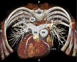
Figure 1. Dual source 64 multi-slice computed tomography volume-rendered image of the heart and the thoracic cage demonstrating the dextrocardia.

AA: ascending aorta; DA: descending aorta; LAD: left anterior descending; LCx: left circumflex; PA: pulmonary artery; RCA: right coronary artery; RI: ramus intermedius; A: anterior; L: left; P: posterior; R: right

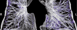
Figure 2. Dual source 64 multi-slice computed tomography volume-rendered image of the lungs depicting the extensive bronchiectasis.
MB: main bronchus; L: left; R: right

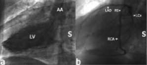
Figure 3. Left heart ventriculography (a) and post-procedural simultaneous left and right coronary arteriography (b) in left anterior oblique projection showing the long axis of the heart.

AA: ascending aorta; LAD: left anterior descending; LCx: left circumflex; LV: left ventricle; RCA: right coronary artery; RI: ramus intermedius; S: spine

Online data supplement

Video 1. Angiography coronaries

Video 2. Angiography left ventricle

Video 3. MSCT coronaries

Video 4. MSCT dextrocardia

Volume 3 Number 2
Aug 20, 2007
Volume 3 Number 2
View full issue


Key metrics

Suggested by Cory

Expert Review

10.4244/EIJ-D-25-00201 Oct 10, 2025
Drug-coated balloons for coronary bifurcation lesions
Fezzi S et al
free

Editorial

10.4244/EIJ-E-25-00040 Oct 6, 2025
Photon-counting computed tomography for stent assessment
Andreini D and Di Mario C
free

Editorial

10.4244/EIJ-E-25-00042 Oct 6, 2025
Seeing is believing
Daemen J and Sadowski K
free

Editorial

10.4244/EIJ-E-25-00044 Oct 6, 2025
Just tap it in... stent optimisation in TAVI
Mylotte D and Thaddeus Soh B
free

Original Research

10.4244/EIJ-D-25-00307 Oct 6, 2025
Impact of clinical risk characteristics on the prognostic value of high-risk plaques
Volleberg R et al

Research Correspondence

10.4244/EIJ-D-25-00139 Oct 6, 2025
In-depth, patient-level analysis of clinical events in the NOTION-2 trial
Khokhar A et al
Trending articles
317.8

State-of-the-Art Review

10.4244/EIJ-D-21-00695 Nov 19, 2021
Transcatheter treatment for tricuspid valve disease
Praz F et al
free
108.3

Viewpoint

10.4244/EIJ-E-22-00007 May 15, 2022
TAVI at 20: how a crazy idea led to a clinical revolution
Eltchaninoff H et al
free
106.35

State-of-the-Art

10.4244/EIJ-D-24-00066 Apr 21, 2025
Management of complications after valvular interventions
Bansal A et al
free
91.6

Image – Interventional flashlight

10.4244/EIJ-D-22-00344 Aug 5, 2022
First dedicated transcatheter leaflet splitting device: the ShortCut device
Tchétché D et al
free
68.05

State-of-the-Art

10.4244/EIJ-D-24-00992 Sep 15, 2025
Antithrombotic therapy in complex percutaneous coronary intervention
Castiello D et al
free
X

PCR
Impact factor: 9.5
2024 Journal Citation Reports®
Science Edition (Clarivate Analytics, 2025)
Online ISSN 1969-6213 - Print ISSN 1774-024X
© 2005-2025 Europa Group - All rights reserved