DOI:

Acute myocardial infarction due to spontaneous coronary artery dissection detected three days before by 64-slice spiral computed tomography

Jiro Aoki*, MD; Gaku Nakazawa, MD; Yoshinobu Onuma, MD; Kengo Tanabe, MD, PhD; Hiroyoshi Nakajima, MD, PhD; Kazuhiro Hara, MD, PhD

A 64-year old man with previous coronary artery bypass grafting was admitted to the hospital with acute ST-elevation myocardial infarction. Three days before admission, multislice computed tomography (MSCT, SOMATOM Sensation Cardiac 64, Siemens Medical System, Forchheim, Germany) was performed due to an ECG abnormality. This revealed inverted T waves in leads V5 and V6 without symptoms. MSCT showed spontaneous coronary dissection in the diagonal branch (Panel 1A through F). Coronary angiography after admission demonstrated total occlusion of the diagonal branch (Panel 2A). Immediate percutaneous coronary intervention was undertaken, and coronary stents were successfully implanted with an excellent final angiogram (Panel 2B). Spontaneous coronary artery dissection is a rare condition that can cause acute myocardial infarction. MSCT is a useful device to detect spontaneous coronary artery dissection before the vessel occlusion.

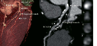
Figure 1. A. Volume-rendered image providing an anatomical overview of the diagonal branch. B. Curved multi-planar reconstructed image of the diagonal branch showing spontaneous coronary artery dissection. The arrowheads indicate cross-sectional images at different levels of the diagonal branch (C, D, E, F). LITA denotes left internal thoracic artery graft. LMCA denotes left main coronary artery. LAD denotes left anterior descending artery. SCAD denotes spontaneous coronary artery dissection.

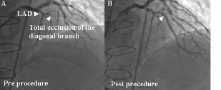
Figure 2. A. Coronary angiogram image of the diagonal branch showing total occlusion. B. Coronary angiogram after the procedure. Two coronary stents were implanted at the diagonal branch.

Volume 2 Number 2
Aug 20, 2006
Volume 2 Number 2
View full issue


Key metrics

Suggested by Cory

Editorial

10.4244/EIJ-D-26-00196 Mar 16, 2026
Is the stress aortic valve index a fractional flow reserve for the stenotic aortic valve?
Kern M and Antoku D
free

State-of-the-Art

10.4244/EIJ-D-25-00512 Mar 16, 2026
Transcatheter aortic valve thrombosis
Jacobsen M et al
free

Original Research

10.4244/EIJ-D-25-01006 Mar 16, 2026
Clinical outcomes and haemodynamic response after blinded stress assessment of moderate aortic stenosis
Eerdekens R et al

Research Correspondence

10.4244/EIJ-D-25-00892 Mar 16, 2026
Extended-length sheaths for TAVI in hostile aortic anatomy
Kobari Y et al

Flashlight

10.4244/EIJ-D-25-00985 Mar 16, 2026
Risk of leaflet avulsion after UNICORN leaflet modification
Khokhar A et al

Flashlight

10.4244/EIJ-D-25-01068 Mar 16, 2026
Three-dimensional intracardiac echo-guided transcatheter tricuspid valve replacement
Swaminathan S et al

Original Research

10.4244/EIJ-D-26-00161 Mar 10, 2026
Long-term Clinical Benefit after Radiofrequency Renal Denervation: Pooled 36-Month Results from the SPYRAL Clinical Program
Kandzari D et al
open access
X

PCR
Impact factor: 9.5
2024 Journal Citation Reports®
Science Edition (Clarivate Analytics, 2025)
Online ISSN 1969-6213 - Print ISSN 1774-024X
© 2005-2026 Europa Group - All rights reserved